Healing Will Take Time & Physical Therapy Can Assist With Recovery

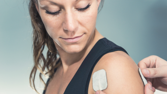
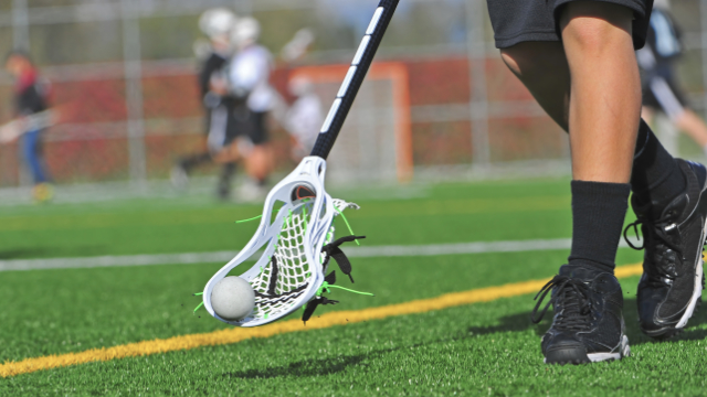
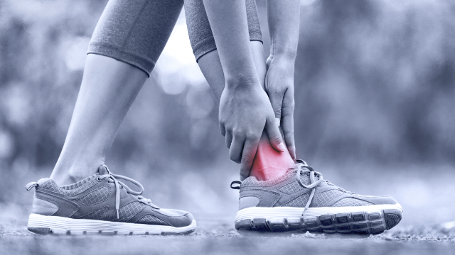
Like it or not, injuries are a part of life. And if you’re an athlete or active individual, this fact is more likely to ring true, as you’re bound to experience an injury from time to time—or perhaps even more often.

After an injury occurs, the question that’s top of mind for most athletes is almost always the same: “how long before I can return?” The response to this question from trainers, physical therapists, and other professionals who work with athletes is often something along the lines of “it’s complicated,” which is accurate due to the various factors and nuances involved in each individual injury. However, there are some general concepts and timelines that can help to give you a better idea of what to expect the next time you get injured.

Average healing times for injured structures

Perhaps the most important concept to understand in this discussion is that although proper rehabilitation can significantly reduce pain levels and help patients regain lost physical function, there are limits to how much the recovery process can be sped up. Injuries cause damage and irritation of one or more structures—sometimes extensively—and it can take a fair amount of time for these structures to repair and remodel afterwards. The amount of blood flow to different tissues and structures also varies widely, which directly affects the amount of time needed for healing. Finally, the severity of an injury will—unsurprisingly—impact healing time, with more severe injuries typically taking longer to heal than mild injuries.

For more context, below is a list of the average time for tissue healing of several commonly injured structures based on a comprehensive literature review:

  • Muscle strain
    • Grade 1: 2–8 weeks
    • Grade 2: 2–4 months
    • Grade 3: 9–12 months
  • Ligament injury
    • Grade 1: 2–8 weeks
    • Grade 2: 2–6 months
    • Grade 3: 9–12 months
    • Surgical repair (eg, ACL): 12+ months
  • Tendon injury
    • Acute: 2–6 weeks
    • Subacute: 2–4 months
    • Chronic: 3–9 months
    • Tear, surgical repair, or rupture: 4–12+ months
  • Other injuries
    • Bone fracture: 6–12+ weeks
    • Articular cartilage: 9–24 months
    • Meniscus/labrum: 3–12 months
  • As you can see, the range for healing times is rather wide for some of these injuries—especially severe ones—which highlights the difficulty of predicting an accurate timetable for returning to sports. Other factors that influence healing time include how much the injured area is loaded, inflammation, cardiovascular health, nutrition, hydration, and sleep, as taking good general care of one’s body can speed up the recovery process. It’s also important to recognize that when pain is no longer detected, it doesn’t necessarily mean that the tissue has completely healed or remodeled. And on the flip side, the presence of pain does not necessarily indicate that there is significant tissue damage. Once again, each injury must be examined on a case–by–case basis by an expert who understands how injuries heal and when it’s safe to return to activity.

    Physical therapy plays a pivotal role in facilitating recovery

    Physical therapists are experts that specialize in helping patients recover from injuries as safely and efficiently as possible. And while we may not have the ability to speed up or change human biology, we do believe that physical therapy can play an essential part in the rehabilitation of most injuries. In each patient encounter we work towards several goals that all contribute to facilitating and expediting injury recovery, including the following:

    • Determine an accurate diagnosis and prognosis
    • Avoid or modify aggravating factors
    • Reduce symptoms, normalize joint motion, minimize swelling
    • Address factors that caused the injury or are making it an ongoing problem
    • Monitor progress and help with exacerbation and recurrences
    • Develop a long–term plan to reduce the risk for injury recurrence

    As physical therapists who deal with injured patients constantly, we understand the frustration of not being given an exact timetable after an injury, but as we’ve shown you, predicting healing times is not an exact science. Injuries are tough, and not knowing when you’ll return is sometimes even tougher. But you should take solace in knowing that a physical therapist will always have your best interests and long–term health in mind, and that all decisions will be based on helping to ensure that you can continue doing what you love long into the future.

    –Summarized from an article published by Evolve Flagstaff

Physical Therapy & Steroid Shots May Both Benefit For Shoulder Pain

Nearly 85% of shoulder conditions involve the rotator cuff, and among the most common of these is shoulder impingement syndrome (SIS). SIS results from the rotator cuff tendons becoming compressed—or “impinged”—as they pass through a small bone on top of the shoulder blade called the acromion. Over time, this causes the tendons to become irritated and inflamed, which eventually leads to bothersome symptoms like swelling and tenderness, loss of strength, restricted movement, and pain.

SIS is most common in individuals that regularly perform lots of overhead activities, especially golfers, swimmers, and baseball and tennis players, as well as painters and construction workers. The condition can also result from an injury that compresses the structures of the shoulder—like a fall—or from frequently sleeping on your side regularly, which can strain the shoulder and cause impingement over time.

SIS is closely related to rotator cuff tendinitis and subacromial pain, and in some cases, the terms are used interchangeably. When any of these conditions develop, the best course of action is a comprehensive physical therapy program, which utilizes movement–based treatments to alleviate pain and increase strength, flexibility, and physical function. However, many patients with SIS go to a primary care physician or some other healthcare provider rather than a physical therapist, and while these providers typically do what they think is best for patients, in some cases they prescribe interventions that are costly and/or ineffective for alleviating shoulder pain.

Researchers review three studies comparing injections to physical therapy

One of these interventions is steroid injections, which some healthcare providers use to provide short–term pain relief for conditions like SIS. However, research analyzing the differences between physical therapy and steroid injections is limited, and this led a team of researchers to conduct a study called a systematic review to compare these two interventions.

For the systematic review, investigators searched a major medical database for studies that compared physical therapy to steroid injections for patients with shoulder pain caused by SIS. This search led to three high–quality studies called randomized–controlled trials being included in the review, which featured data on 452 patients.

Results from these three studies revealed that both physical therapy and steroid injections led to improvements in pain, shoulder range of motion (ROM), and shoulder function in the short term (1–3 months), medium term (6 months), and long term (12 months). Although steroid injections were more effective than physical therapy for improving shoulder function in the short term at 6–7 weeks, there were no significant differences between these interventions in pain, shoulder ROM, or shoulder function at the medium–term or long–term follow–ups.

Based on these findings, it appears that both physical therapy and steroid injections provide similar benefits for patients with shoulder pain caused by SIS, with neither intervention found to be superior to the other. Therefore, researchers recommend that patients should be educated on the risks versus benefits of each intervention and given the choice between interventions based on their preference. As a part of this discussion, patients should be informed that physical therapy is generally regarded as a lower–risk treatment option, as evidence on the long–term risks of steroid injections is limited and some studies have found that injections can lead to a compromised immune system and cartilage toxicity over time.

If you’re currently dealing with SIS, we encourage you to explore all treatment options available to you before making a decision. And if you decide that physical therapy is right for you, we’d be more than happy to get you started on a comprehensive treatment program right away.

Having an MRI Unnecessarily May Increase Healthcare Costs

Low back pain is jarringly common. About one–half of all working Americans experience symptoms at least once every year, and roughly 31 million are affected by it at any given point in time. So if you happen to place yourself in this category, you’ll have an abundance of company.

Dealing with low back pain can be troublesome and place a strain on everyday life. Typical movements like bending over to pick something off the ground or twisting your torso when looking to the side might suddenly make you pause and cause you to be less mobile as a result. This naturally leads to frustration and can often shift to a focus on one main question: “what’s causing my pain?”

Many patients with low back pain therefore begin to place a strong emphasis on obtaining a diagnosis. Patients who do this usually believe that obtaining a diagnosis will clearly explain what’s causing their pain and will make it easier for them to receive appropriate treatments. But sadly, searching for a low back pain diagnosis is complicated and often does not lead to the outcomes that most patients hope for. And in many cases, it can do more harm than good.

When seeking a diagnosis, many patients will have an imaging test (X–ray, MRI, or CT scan) performed, either by the doctor’s order or their request. These types of tests are essential for diagnosing numerous conditions throughout the body, but when it comes to low back pain, their usefulness is limited. The primary issue is that imaging tests are only one component of a diagnosis, in addition to a detailed patient interview and thorough physical examination. Plus, many “abnormal” results from imaging tests could be simple age–related changes that are not contributing to a patient’s pain, but this won’t stop certain practitioners from treating the “problem” nonetheless.

Clinical guidelines and experts have long recommend that imaging tests for low back pain should only be performed if one or more “red flags” is identified during an examination. Red flags for low back pain include the following:

  • Loss of bladder or bowel control
  • Signs of severe or worsening nerve damage
  • Serious underlying problems like cancer or spinal infections
  • Unexplained weight loss
  • Abnormal reflexes
  • Recent serious fall or injury
  • Worsening numbness or weakness in one leg

If none of these red flags are present, having an imaging test is not recommended because it’s not likely to provide any valuable information or lead to better outcomes. Yet many patients with low back pain and no red flags still undergo MRIs of their spine.

Greater use of healthcare services leads to higher costs for patients undergoing MRIs

A 2015 study illustrates the implications of unnecessarily having an MRI for low back pain instead of seeing a physical therapist. For the study, researchers analyzed data from the medical records of 2,893 patients with low back pain that were identified through a comprehensive search. Of these patients, 841 received treatment outside of primary care within the first six weeks of their diagnosis, with 46% receiving a diagnostic test—usually an MRI—and 45% receiving physical therapy.

A comparison of these two groups showed that those who received a diagnostic test first utilized significantly more healthcare services than those who underwent physical therapy first. For example, patients who first received a diagnostic test were more than 3 times more likely to undergo surgery, almost 4 times more likely to have injections, and about 7 times more likely to see a spine surgeon compared to those who saw a physical therapist first. As a result, healthcare costs for low back pain over one year were about $4,700 higher when imaging was performed first due to the increased use of these healthcare services.

This study clearly shows why it’s usually best for patients with low back to visit a physical therapist early after noticing pain rather than going to a primary care physician or specialist, who may be more likely to order a diagnostic test. With this in mind, we strongly recommend that you consider seeing a physical therapist if low back pain is bothering you and to avoid the temptation of relying too heavily on a diagnosis for the reasons we’ve described here.

Physical Therapy May Be The Best Option for Knee Osteoarthritis

Knee osteoarthritis is a disorder that involves age–related changes to the cartilage in a knee joint. In a normal knee, the ends of each bone are covered by cartilage, a smooth substance that protects the bones from one another and absorbs shock during impact. In knee osteoarthritis, this cartilage becomes stiff and loses its elasticity, which makes it more vulnerable to damage. Cartilage may begin to wear away over time, which greatly reduces its ability to absorb shock and increases the chances that bones will touch one another. When this occurs, it typically results in pain within and around the knee that gets worse with activities like walking, going up/down stairs, or sitting/standing. Swelling, tenderness, and stiffness are also common.

Although no treatment can slow or stop this loss of cartilage, physical therapy is strongly recommended as an initial intervention for all cases of knee osteoarthritis. Undergoing a comprehensive course of physical therapy can help reduce pain levels and preserve knee function through movement–based strategies like stretching and strengthening exercises, hands–on therapy, bracing, and recommendations on how to modify pain–inducing activities. Physical therapy can also reduce the need for other interventions that may be potentially unnecessary or dangerous, such as surgery or opioids, and research has shown that the earlier physical therapy is initiated, the greater its benefits.

Researchers review data over 20 years to search for connections between early physical therapy and opioid use

Despite these recommendations, many patients with knee osteoarthritis either never see a physical therapist or fail to do so until much time has passed, which can lead to worse outcomes. With this in mind, a study was conducted to investigate whether there is an association between early versus late initiation of physical therapy for knee osteoarthritis and the future use of opioids.

For the study, investigators searched Medicare and commercial health insurance claims data from 1999 to 2018 for information on adults with knee osteoarthritis who were referred to physical therapy within one year of their diagnosis. Patients identified through this search were then categorized as either “opioid naïve” (meaning they did not use opioids) or “opioid experienced” based on prescription history with these drugs before initiating physical therapy for knee osteoarthritis. Finally, researchers examined the relationship between when physical therapy was initiated and with the use of opioids—including chronic opioid use—over 1 year.

Researchers identified 67,245 patients with knee osteoarthritis, 35,899 of whom were classified as “opioid naïve” and 31,346 of whom were “opioid experienced.” In the opioid naïve group, the risk for any opioid use was higher for patients that delayed starting physical therapy compared to those who began within one month of their diagnosis, and this risk continued to increase as the length of the delay grew (up to 12 months). Similar results were found for the risk of chronic opioid use, which was 2.5 times higher for patients who waited 9–12 months to see a physical therapist compared to those who started physical therapy within one month. The same trends were identified in the opioid experienced group, with increased risks for both opioid use overall and chronic opioid use in patients who waited to see a physical therapist versus those who saw one within one month.

These findings suggest that delaying the start of physical therapy may increase the risk for using opioids compared to starting it early (within one month) for patients with knee osteoarthritis, and the longer the delay, the greater the risk for opioid use. Therefore, if you’re currently dealing with knee osteoarthritis, we strongly recommend that you visit a physical therapist sooner rather than later.

Adequate Sleep Is Integral To Athletic Recovery

We spend roughly one–third of our lives sleeping, so it follows that the habits we keep during the night have a major impact on our waking lives. Sufficient sleep is an essential component of good overall health, as getting between 7–9 hours of sleep every night is linked with countless benefits, including stress relief, a reduced risk for many chronic disorders, improved memory and cognitive function, and possibly weight loss and a longer lifespan. Proper sleep is even more essential for athletes and anyone dealing with a painful condition, as it represents one of the most effective recovery strategies one can follow. Some experts even recommend that athletes should aim to get 9–10 hours of sleep each night to help them reach their full potential.

To elaborate on the association between sleeping habits and recovery, here are a few research examples that highlight the negative impact of sleep deprivation on both physical and cognitive performance:

  • Individuals with poor sleeping habits have lower general health and increased stress and confusion
  • Inadequate sleep impairs maximal muscle strength during certain movements
  • Adolescent athletes who average less than 8 hours of sleep per night have a 1.7 times greater risk of injury than those who sleep more than 8 hours per night
  • Sleep may be important for bone health, and sleep deprivation may contribute to the development of bone stress injuries
  • Insufficient sleep and chronic pain have a bi–directional relationship, meaning they each contribute to one another; one study found that <7 hours of sleep was associated with a significant increase in the risk for new injuries in athletes, while getting >7 hours was linked to a significant decrease in injury risk

Yet despite this growing body of evidence, many individuals still aren’t getting enough sleep. Statistics suggest that 35% of Americans get less than 7 hours of sleep per night and 63% claim that their sleep needs are not met during the week. This problem exists in athletic populations as well, with research showing that 50–78% of elite athletes experience sleep disturbance and 22%–26% suffer from highly disturbed sleep. The reasons why so many don’t get adequate sleep are complex and multifaceted, but some contributing factors include the fast pace of modern life, dietary habits, insomnia, and excessive light exposure from bright screens—especially at nighttime—which suppresses melatonin, the major hormone that controls sleep and wake cycles.

Take better control of your sleeping habits with these tips

If you’re trying to improve your sleeping habits, here are some of the most effective strategies to help you make it happen:

  • Spend as much time as possible outdoors and exercise regularly
  • Make your bedroom a sanctuary and keep it cool, quiet, and dark, with a high–quality mattress that’s comfortable and not worn out
  • Make sure your final hour before bed is relaxing and free of much stimulation; avoid bright screens during this time, as well as eating, working, or reading in bed
  • Try to go to bed and wake up around the same time every night; on weekends, try to stick to this and only allow about a one–hour difference
  • Avoid nicotine and caffeine, especially in the final hours before bed, since they are stimulants that can interfere with sleep
  • Avoid big meals a few hours before and alcohol right before bedtime
  • If needed, squeeze in a nap during the day, but try to keep it to 20–30 minutes and only take them in the early afternoon

It’s also essential to identify and remove any barriers that might be directly interfering with proper sleep. For example, if you’re dealing with pain that’s preventing you from getting enough sleep, you need to first take steps to address it—like seeing a physical therapist—before you can expect the other strategies to work effectively. Self–reported measures of sleep like sleep diaries and fitness trackers that monitor sleep can be extremely helpful as well. And for athletes, an additional step is to work with coaches and other team members to discuss your changing sleep habits and collectively identify approaches that work for you.

The importance of sleep cannot be overstated, and even getting slightly more sleep than you’re currently getting can make a big difference. We understand that it’s easy to neglect sleep in an age when life moves fast and distractions seem to be everywhere, but if you take your sleep seriously, you’ll likely start to notice significant changes in your athletic performance and life in general.

TENS Therapy Provides Immediate, Short-Term Pain Relief

Physical therapists utilize a variety of interventions for each patient they treat, and their guiding principle is to always combine whichever interventions are most likely to produce a successful outcome. Most treatment programs will include a combination of exercises to increase strength and flexibility, hands–on manual techniques to alleviate pain and improve function, and passive modalities that are also used to reduce pain levels.

One of the more commonly used modalities is called transcutaneous electrical nerve stimulation (TENS), which is a noninvasive therapy that sends a low–voltage electrical current through the nerves to alleviate pain. As a noninvasive treatment, TENS can be seen as an alternative to drug therapy that comes with far fewer side effects. Compared with pain–relieving drugs, the most common side effect of TENS therapy is an allergic skin reaction, which only occurs in about 2–3% of patients.

TENS is administered with a small, battery–powered machine about the size of a pocket radio. The TENS machine has two wires that conduct electrical current called electrodes, which are attached to the skin at the area where the patient is experiencing pain. This creates a circuit of electrical impulses that travels along nerve fibers to relieve pain in that region.

When the TENS machine is turned on, it can be set for different wavelength frequencies depending on the location and degree of the patient’s pain. Once the current is delivered, the patient will often notice an immediate reduction of their pain level. This is either because the electrodes stimulate the nerves in an affected area and signals to the brain that block or scramble normal pain signals, or because the electric stimulation of nerves may help the body produce natural pain killers called endorphins, which block the perception of pain.

Large–scale study confirms the efficacy of TENS therapy for pain

Although TENS therapy is used frequently, its efficacy in the real world has been a matter of uncertainty and controversy for many years. Therefore, a large–scale study called a systematic review and meta–analysis was conducted to investigate the efficacy and safety of TENS therapy.

For the meta–analysis, researchers performed a search of 10 major medical databases for high–quality studies that evaluated the use of TENS versus either no treatment, placebo, or some other treatment, for adult patients with clinical pain. This search led to 381 studies being included, with 91 studies comparing TENS to placebo, 10 comparing TENS to no treatment, 61 comparing TENS to standard of care treatments, 67 comparing TENS to other treatments, and 13 comparing high–frequency to low–frequency TENS.

Results from the meta–analysis revealed that there was moderate–certainty evidence that TENS therapy applied within or close to the site of pain elicits clinically important reductions in the intensity of pain during or immediately after treatment. In addition, there were no reports of serious adverse events, which shows that TENS is can be safely used to address pain.

According to researchers, these findings are consistent with clinical experience and long–held expert opinion that TENS provides some patients with immediate short–term pain relief. Researchers also claim that this study should resolve long–term uncertainty about the efficacy of TENS and should encourage physical therapists and other healthcare providers to consider using TENS as an adjunct to core treatment for immediate short–term relief of pain for most musculoskeletal conditions. In addition, patients who are treated with TENS are advised to tailor treatment according to their individual needs to maximize the benefit of this modality.

The Use of Walking Aids Is Essential To Reduce The Risk Of Falling

If you’re over the age of 75, you might worry about the possibility of falling on a regular basis. This concern is completely understandable, as falls are the leading cause of injury for older adults, and the likelihood of falling increases with each additional year of age. About 40% of older adults living at home experience a fall once per year, and this figure is even higher for adults living in nursing homes and assisted living facilities.

One commonly recommended strategy to mitigate the risk of falling is for older adults to use a walking aid like a cane or front–wheeled walker. These devices assist with upright balance and mobility, which are essential for avoiding a fall; however, some research has suggested that surprisingly, using a walking aid may be an independent risk factor for falling. This is likely due to lack of guidance on how to use these devices, as one study found that over 80% of wheeled frame users reported not receiving any instructions on how to use it and another found that 66% of patients with hip problems were not educated on which hand to hold their assistive device in.

In response, a 2020 study was conducted to investigate older adults’ use of walking aids, how their usage patterns affect stability, and what the general perception is of walking aids from the perspective of users and healthcare professionals.

17 older adults are closely monitored while using a walking aid

For the study, researchers identified 17 older adults with an average age of 70 that regularly used a walking aid. These participants then completed the Falls Efficacy Scale—which measures concerns about falling—and were evaluated in different environments several outcome measures. They were also recorded on video while using a front–wheeled walker to assess their techniques. Finally, two focus groups were formed, with one comprised of 5 walking aid users and the other comprised of 10 healthcare professionals. These focus groups were intended to stimulate meaningful discourse about the use of walking aids and identify any areas that could be improved.

Results showed that the percentage of time in which the walking aid was used incorrectly varied between participants, but all participants displayed incorrect usage at some point in the study. On average, the group had incorrect usage of walking aids during 16% of their single support periods and 30% of dual support periods. Video analysis also showed that many participants did not use front–wheeled walkers properly. The most common error was lifting the frame when turning, which is risky and can lead to a fall.

The walking aid user focus group identified several key themes, one of which was that participants considered the lack of guidance in the safe use of walking aids to be a problem. Members of the healthcare provider focus group agreed that a gold–standard, universally accepted approach was lacking for the prescription of walking aids and training of users, which highlights a gap that needs to be addressed. Healthcare providers reported that they instead rely on their clinical judgement regarding the user’s safety and gait performance, and that they train patients on how to use a walking aid based on practice and repetition rather than established guidelines.

Overall, results suggested that adherence to current guidance on how to safely use a walking aid in the home is poor amongst users, and that both environmental and design–related factors serve as barriers to adherence. Participants also displayed low stability during incorrect use of walking aids, which indicates that they are putting themselves at risk of falling when improperly using these devices in various environments.

Key tips to ensure safe use of walking aids

These findings highlight the importance of proper use of walking aids and the need for better training of walking aid users. To accomplish this, experts must work together to establish clearer guidelines for healthcare providers who train patients to use walking aids, but in the meantime, here are a few key tips to help ensure you’re using a front–wheeled walker (a popular type of walking aid) properly:

  • The height of the walker should be adjusted so that your arms are slightly bent (about 20–30 degrees) when using it
  • When using a front–wheel walker, be sure to keep the front of your body in line with the back two posts of the walker
  • Advance the walker a few inches in front of you first, and make sure all tips and wheels are touching the ground before taking a step
    • When you’re ready to take a step, step forward with your bad leg first, followed by your good leg, placing it in front of your lead foot
  • When turning the walker, keep the walker in front of you and take small steps; DO NOT pick the walker up at any time
  • Don’t place all your weight on the walker; allow it to assist your gait by bearing only part of your weight upon it
  • Add tennis balls to the bottom of the walker to make it glide more smoothly

If you or someone you care for uses a walking aid, we can provide additional guidance on how to properly use these devices to reduce the risk for falling. Contact us for more information.

Seeing A Physical Therapist Can Help Ensure A Safe Return To Sports

In the realm of sports–related injuries, the hamstring is one that most people are at least somewhat familiar with. This makes sense, as a pulled hamstring—or hamstring strain—is one of the more common injuries in sports. The pain and movement restrictions that result from this type of injury usually prevents an athlete from participating in their respective sport for some time, but following a course of physical therapy can lead to a fast recovery and safe return to sports for patients dealing with a hamstring strain.

Although it might be assumed to be a single muscle by some, the hamstring is a muscle group consisting of three separate muscles—the semitendinosus, semimembranosus, and biceps femoris—that run down the back of the thigh from the lower part of the pelvis to the back of the shinbone. These three muscles serve a crucial role, as they help the knee joint to bend (flex) and the hip joint to straighten (extend). The hamstring is balanced by the quadriceps muscles in the front of the thigh, which are responsible for the opposite function: extending the leg at the knee joint and flexing the thigh at the hip joint. Together, they control the power and stability of the knee joint and allow for running and other activities.

A hamstring strain can involve any of the three hamstring muscles, and the most common cause for an injury is overloading one or more of these muscles. This can occur when a muscle is stretched beyond its capacity or challenged with a sudden load, which is why hamstring strains are so common in sports, especially those that involve lots of sprinting or sudden changes in direction. Therefore, athletes who participate in basketball, football, tennis, soccer, and the sprinting events of track—as well as dancers and gymnasts—have the highest risk for sustaining a hamstring strain. Other risk factors include prior hamstring injury, muscle tightness, poor conditioning, and older age.

When a hamstring strain occurs, the most common symptom is a sharp pain in the back of the thigh that may be felt immediately. Patients may also feel a “popping” or tearing sensation in this region, which may be followed by tenderness or swelling that may develop within a few hours. Some patients may experience bruising or discoloration on the back of the thigh or persistent weakness in this area as well.

Physical therapists always ensure patients are ready to return to sports

A hamstring strain may sound like a daunting injury, but for most patients, conservative (non–surgical) treatment and some time away from sports is all that’s needed. Physical therapists are uniquely positioned to manage patients with hamstring strains and can help them recover quickly and safely through individualized and evidence–based treatment. A standard treatment program for a patient with a hamstring strain will consist of the following:

  • Range of motion exercises: once initial pain and swelling have subsided, the patient will start with some gentle stretching exercises like a hamstring stretch to improve flexibility
  • Strengthening exercises: includes various strengthening exercises to build back strength in any areas that are weak
  • Manual therapy: involves the therapist applying hands–on techniques to the patient’s muscles and joints to alleviate pain and improve range of motion and strength
  • Functional training: this type of training includes exercises that are specifically catered to the sport or activity that the patient is returning to

Another significant benefit of having physical therapy for a hamstring strain is that therapists always take every measure to ensure that athletes don’t return to sports until they have completed their rehabilitation and can do so with a minimal risk for injury. This is accomplished by structuring the timeline of programs based on the average time needed to recover from a hamstring strain, closely evaluating the athlete’s progress along the way, and then assessing the athlete when they are nearing a return to ensure they fulfill all appropriate criteria. Only then will the therapist provide the go–ahead that the athlete can safely return to his or her sport.

According to a proposed algorithm for hamstring strains, an athlete will be ready to return to their sport once they meet the following criteria:

  • No tenderness when pressure is applied to the hamstring
  • Adequate muscle strength based on strength testing
  • No insecurity on the active hamstring test, which is done by performing a straight leg raise as fast as possible to the highest point without fear of injury
  • Ability to complete 30 or more repetitions of the single leg bridge test
  • No pain or hesitation with sport–specific movement testing (eg, accelerations, decelerations, rotations, sprinting, cutting, pivoting, jumping, or hopping)

So if you’ve recently injured yourself and your symptoms sound similar to a hamstring strain, we strongly recommend seeing a physical therapist to help you get started on your path to recovery.

Study Identifies Two Metals In Many Popular Chocolate Brands

If you have a tough time ending your day without reaching for a piece of chocolate—or four—to satisfy your sweet tooth and give you that blissful boost that few other foods can provide, you might want to up. Although dark chocolate certainly tastes good and may provide several health benefits due to its high concentration of antioxidants—which can improve heart health and other conditions—a recent study has shown that several dark chocolate bars contain lead and cadmium, two heavy metals that have been linked to a variety of health conditions.

Chocolate makers are aware of this issue for some time and have been trying to find ways to reduce the levels of these metals for some time, but it’s been unclear whether they’ve succeeded. Therefore, scientists at Consumer Reports performed a study in which they measured the amounts of heavy metals in 28 dark chocolate bars, and the results were less than encouraging.

The scientists tested various brands of dark chocolate bars, including lesser–known brands as well as popular brands like Dove and Ghirardelli. They found lead and cadmium to be present in all 28 bars, and for 23 of these bars, eating just one ounce of chocolate per day would put an adult over the level that experts say can be harmful to one’s health. Consistent, long–term exposure to heavy metals like lead and cadmium can lead to numerous health conditions, such as nervous system issues, hypertension, immune system suppression, and kidney damage. The risk for health problems is highest in pregnant women and young children, as exposure to these metals can affect brain development and cause developmental delays.

Lead and cadmium? How did they get in my chocolate?

As you’re processing the—probable—shock of reading that your favorite dark chocolate bar likely contains unhealthy levels of lead or cadmium, your next question might be: why are these metals found in chocolate?

Chocolate is made from the cacao bean, which is composed of cocoa solids and cocoa butter, which are together called cacao or cocoa. Dark chocolates are generally at least 65% cacao by weight, which means they have high concentrations of cocoa solids. These cocoa solids are where the nutritional value of dark chocolate is derived, since they are packed with antioxidants called flavanols that are associated with better blood vessel function and lower cholesterol and inflammation. But the cocoa solids are also where heavy metals like lead and cadmium are found, which is why levels are so high in dark chocolate.

These two metals get into chocolate through different mechanisms. Cacao plants take up cadmium from the soil and the metal accumulates in the cacao beans as the tree grows, while lead appears to enter cacao after the beans are removed from pods and dried out, during which time lead–filled dust and dirt accumulates on the outer shells of the beans. But because of these different entry methods, the techniques used to reduce lead and cadmium levels are also different. For lead, changes are needed in harvesting and manufacturing practices to minimize the amount of lead–contaminated dust that lands on gets into the beans as they’re processed, while reducing cadmium levels is more difficult and may require carefully breeding or genetically engineering plants to take up fewer heavy metals, which will take much longer. Chocolate producers can also survey the areas in which they grow cacao and favor beans grown from regions with lower levels, or blend beans from high–level areas with lower–level areas.

And for you, the consumer, here are some tips to help you navigate this new bombshell on your favorite treat:

  • Choose dark chocolates with the lowest levels of heavy metals (see the Consumer Reports article for more details)
  • Limit your chocolate intake to only a few days per week, at most
  • Try dark chocolates with lower cacao percentages (eg, 65–70%), and consider alternating it with milk chocolate (but remember that milk chocolate is higher in sugar)
  • Don’t give kids much dark chocolate
  • Eat a well–rounded diet that’s high in whole grains and fruits and vegetables, and low in processed foods and those that are high in refined carbs and sugars

The long and short here: there’s no need to ditch your chocolate habit altogether because of these findings, but you should reevaluate your chocolate intake and make necessary changes to ensure that you’re limiting the amount of lead and cadmium you’re consuming.

Rehabilitation Helps Athletes Return To Their Sport Quickly & Safely

About 25,000 people sprain their ankle every day, and in most of these incidents, sports are involved. Ankle sprains represent the most common injury in sports, as they account for a whopping 45% of all sports–related injuries. But this risk varies widely between sports, with football, basketball, and soccer being associated with the highest rates of ankle sprains because they involve high speeds and frequent changes in direction. In football, for example, ankle sprains occur at a rate of 1/1,000 hours, meaning that one ankle sprain occurs for every 1,000 hours of participation.

These statistics may seem daunting, but a recently published review has shown, most ankle sprains can be effectively treated with a conservative treatment program that involves evidence–based rehabilitation.

Ankle anatomy and grading system

Ankle sprains involve the ligaments of the ankle joint, which are flexible bands of tissue that connect one bone to another. Ligaments are elastic and can be stretched to a certain length and then return to their original position, but they have a limit. When any ankle ligament is stretched beyond its maximum range of motion, damage will occur, and the result is an ankle sprain. Ankle sprains are generally categorized into the following three groups:

  • Grade 1 (mild): ligament(s) stretched but there is no tear; symptoms involve mild pain and tenderness, some swelling and stiffness
  • Grade 2 (moderate): most common type of sprain; ligament(s) partially torn; symptoms include significant swelling and bruising, moderate pain, and trouble walking
  • Grade 3 (severe): ligament(s) completely torn; symptoms involve severe swelling and pain, especially while walking, instability of joint, extreme loss of motion, possible difficulty bearing weight on foot

Depending on its location in the ankle, a sprain can be further categorized as either lateral, medial, or high. Lateral ankle sprains take place on the outside part of the ankle, which is the most common site for a sprain (about 80% of all sprains). High ankle sprains are less common (up to 15% of sprains) and are often seen in football, downhill skiing, and other field sports, while medial sprains are the least common (about 6%).

Most patients will make a complete recovery after sustaining an initial ankle sprain, but up to 70% of those who experience a lateral ankle sprain will go on to develop chronic ankle instability. Patients with this condition experience changes in the function of their nervous system that may lead to decreased postural control, joint position awareness, and more ankle instability. The combined result of these changes is an increased risk for recurring pain and other symptoms, as well as greater odds for sustaining a second ankle. And with each additional sprain, the risk continues to rise.

Appropriate rehabilitation is key to successful outcomes

This underlines the importance of proper treatment after the first ankle sprain, which can significantly reduce the risk for chronic ankle instability. Fortunately, most ankle sprains can be effectively treated with conservative (nonsurgical) interventions, and patients can expect to experience good to excellent outcomes when following this approach. One of the only possible exceptions is grade 3 lateral ankle sprains, as surgery may be beneficial for some elite athletes dealing with this injury to accelerate their recovery; however, conservative treatment is still preferred over surgery in most cases.

Physical therapy is generally regarded as the best way to deliver conservative treatment to patients, since therapists utilize individualized and evidence–based rehabilitation programs to achieve the highest level of care. Ankle sprain rehabilitation typically begins with alleviating swelling and further injury during the initial inflammatory phase of recovery, which is achieved with the POLICE (protect, optimal loading, ice, compression, elevation) protocol for the first 2–7 days. Short–term immobilization with a removable cast or boot may also be helpful for severe ankle sprains during this period. After inflammation has subsided, patients are advised to wear an ankle brace and will begin an exercise therapy program, which typically includes early active range of motion exercises, followed by strengthening exercises, proprioceptive training, and functional exercises. Exercises should simulate the physical demands of the patient’s sport and become more challenging as the program progresses.

After an ankle sprain, the top concern of most patients is when they will be able to return to their respective sport. But unfortunately, this decision is often difficult, as returning a player too soon can lead to residual disability and additional injuries in the future. Therefore, it is the physical therapist’s responsibility to ensure that athletes do not return too soon by ensuring that certain functional markers are met.

There are currently no formal criteria to clearly determine when an athlete is ready to return to sports, but several tests can be used to assist in this decision. When analyzing patients, therapists must ensure that all functional limitations from the sprain have been restored, cardiovascular fitness is equal to or greater than pre–injury status, and that there is no apprehension from the athlete or other members of the rehabilitation team concerning their safety. For patients with a history of ankle sprain, extra caution is required since the risk for chronic ankle instability is higher.

All patients must also recognize that although an extended recovery can be frustrating, taking the adequate time to heal and recover will increase their chances of long–term success. We will work to manage your expectations and ensure that you’re getting back to the field or court as quickly, but also as safely, as possible