The downsides of overusing diagnostic tests

As we explained in our last newsletter, diagnostic imaging tests can be a vital tool that helps doctors more clearly understand what’s happening in the body in order to reach or confirm a diagnosis. But simply because these tests are helpful does not mean they should be used at all times, or even frequently. For musculoskeletal disorders—like back, neck, and knee pain—imaging tests should be used when certain criteria are met, yet statistics on their use paint a much different picture.

The overuse of diagnostic testing for musculoskeletal disorders is part of a much larger trend in the healthcare industry. According to one article, about $800 billion of healthcare spending is spent on unnecessary diagnostic tests, procedures, and extra days in the hospital. For musculoskeletal disorders, back pain typically garners the most attention because it is so common and because it serves as one of the clearest examples of excessive diagnostic testing use.

Low back pain is one of the most common overall ailments and will affect up to 80% of individuals at some point in their life. Imaging tests are only recommended when a “red flag” is present, which include signs of severe or worsening nerve damage, serious underlying problems like cancer or spinal infections, unexplained weight loss, loss of bladder or bowel control, and abnormal reflexes. If none of these red flags are present, an imaging test is not usually required, but many patients with back pain continue to have MRIs of their spine for one reason or another.

Despite the existence of guidelines that recommend against MRIs, except for in these rare cases, there is data that shows many patients with back pain are initially being prescribed diagnostic imaging tests without first attempting less expensive and less invasive treatments like physical therapy. In fact, one study estimated that the use of MRIs for low back pain increased by as much as 300% between 1994-2006. And while it may seem as if the overuse of these helpful tests is generally harmless, the inverse is actually true.

For starters, imaging tests—particularly MRIs and CT scans—are extremely expensive. The average cost of an MRI is $1,119, with some costing as much as $3,000, which is significantly higher than any other comparable countries. CT scans and X-rays also expose patients to potentially harmful levels of radiation that could otherwise be avoided. But perhaps most importantly, imaging tests can do more harm than good for many patients. This is because they don’t always identify the cause of patients’ pain—especially for low back pain—and often reveal “abnormalities” unrelated to the pain that may be misconstrued as a cause. An important study highlights why this is true, as it reviewed the MRIs and CT scans of more than 3,000 individuals with no signs of back pain. Results revealed the following:

  • 20-year-olds: 37% had “disc degeneration” and 30% had “disc bulging”
  • 50-year-olds: 80% had “disc degeneration” and 60% had “disc bulging”
  • 80-year-olds: 96% had “disc degeneration” and 84% had “disc bulging”

These results show that disc degeneration and disc bulging are extremely common in most people without back pain, and the likelihood of having these “abnormalities” increases significantly with age. Therefore, many patients with back pain may be told that they have disc degeneration or disc bulging and believe this to be the reason for their pain, while the truth is that it is likely a sign of the natural aging process instead.

Another extremely unfortunate consequence of this testing is that many patients go on to receive invasive, dangerous, and expensive interventions based on the results of their tests, even if the procedure won’t actually resolve their pain. In our next newsletter, we’ll explore the repercussions of overusing diagnostic imaging tests in more detail and explain why this can all be avoided by seeing a physical therapist first.

What is diagnostic imaging testing and when is it needed?

When dealing with pain of any sort, physical therapists and medical doctors have a number of tools at their disposal to help determine the cause and what needs to be done to address it. A thorough physical examination that involves a variety of objective and subjective assessments of strength, flexibility, balance, and other variables is always the first and most important step of reaching an accurate diagnosis. But in some cases, additional testing may also be performed.

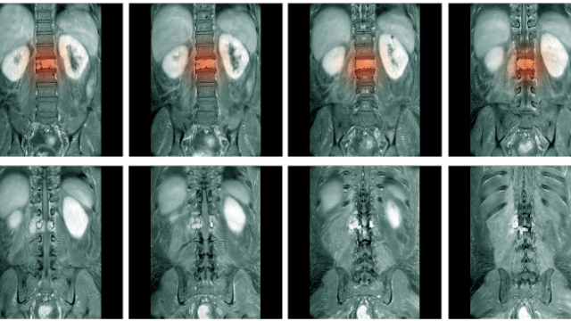
If your doctor is still uncertain of what is causing your pain or if a severe injury was sustained, she may recommend having a diagnostic imaging test to obtain more information. These non–invasive techniques let the doctor see inside your body to get a clearer picture of your bones, muscles, tendons, and ligaments. This can help to determine if any of these structures look damaged and are possibly contributing to your pain. Diagnostic imaging tests include x–rays, computed tomography (CT) scans, and magnetic resonance imaging (MRI) scans, which all use slightly different methods to produce images of the internal structures of the body.

  • X–rays
    • The most commonly used diagnostic test; readily available in many doctor’s offices
    • Sends a type of electromagnetic radiation called X–rays through the body
    • Bones and other dense matter appear white or light because they absorb the radiation, while less dense soft tissues (like ligaments and tendons) and bone fractures look darker because they let radiation pass–through
    • X–rays do not show as much detail as more sophisticated tests but are often used as a starting point
    • You are exposed to radiation which can, in some cases, cause harmful effects to cells of the body
  • CT scan
    • Combines X–rays with computer technology to produce a more detailed image that includes the size, shape, and position of structures deep in the body
    • During the test, an X–ray tube will rotate slowly around you and take several pictures from all directions, which are displayed on a computer screen
    • May be needed for problems with small, bony structures or severe trauma
    • You are exposed to a much higher dose of radiation than with an x–ray, which in some cases, can cause harmful effects to cells of the body
    • Costs more and takes more time than regular X–rays
  • MRI
    • Uses magnetic fields and computerized technology instead of radiation to take high–resolution pictures of bones and soft tissue
    • Involves lying on a table that slides into the MRI scanner. MRIs employ powerful magnets which produce a strong magnetic field that forces protons in the body to align with that field. When a radiofrequency current is then pulsed through the patient, the protons are stimulated, and spin out of equilibrium, straining against the pull of the magnetic field; a computer records how these tissues respond to these radio pulses and then translates the data into a detailed picture
    • May be used in helping to diagnose torn ligaments and cartilage, torn rotator cuffs, herniated disks, osteonecrosis, bone tumors, and other problems
  • Other
    • Ultrasound: is a radiation–free technique that uses high–frequency sound waves that echo off body structures to diagnose a variety of conditions
    • Bone scan: shows bone activity throughout the body by injecting a small amount of radioactive material into a vein, which is absorbed by areas forming new bone (like fractures and bone tumors) and can be identified by a scan of the body several hours later

Each of these techniques can serve a unique and important role in the diagnosis of many conditions, but there are only certain situations in which they should be used. In our next newsletter, we’ll look into why diagnostic imaging tests are being overused and the downsides of this ongoing trend.

The best way to recover from a sports injury is with physical therapy

As we explained in our first newsletter this month, sports injuries are unfortunate but also a common occurrence for athletes. A range of factors can contribute to an athlete’s risk for injury, including their participation in a higher level of activity without first preparing through a tailored training program.

Returning to any sport after an extended absence can be dangerous no matter what physical condition you happen to be in. The reason is that even if you’re in good shape, your body may not be prepared for the particular dynamics involved in the sport. Every sport requires a very specific set of movements, and in order to perform them, specific areas of the body need to be appropriately prepared with proper strength and flexibility. Overworking any muscle groups or ligaments that are not yet adapted to these movements can result in damage to these structures and injury.

The pandemic has temporarily suspended most organized sports, because of this there is likely to be an uptick in sports injuries when things return to normal circumstances. Without organized sports functioning now, many individuals have not been keeping up with the same level of training as they had before the pandemic. If they jump back into their sport too aggressively, it could prove to have a dangerous outcome. Below are some of the most common sports–related injuries that may be seen frequently during the return to the sporting world ahead:

  • Baseball: sprains and tears of the ulnar collateral ligament, Little League elbow and Little League shoulder
  • Softball: shoulder tendinitis, back and/or neck pain
  • Tennis: tennis elbow, rotator cuff injuries
  • Golf: back pain, golfer’s elbow, shoulder pain
  • Track & field/running: runner’s knee, jumper’s knee, IT band syndrome
  • Swimming: swimmer’s shoulder, hip pain, back pain
  • Lacrosse: ACL tears, muscle strains
  • Soccer: strains and sprains to the lower leg, especially the ACL

Why you should see a physical therapist first and fast

If an injury does occur, the smartest and safest decision you can make is to see a physical therapist right away. Physical therapists are movement experts with a thorough understanding of the biomechanics involved in all sports. As such, they can help patients prepare for the demands of their respective sport to prevent injury, and if an injury does occur, guide them through a comprehensive rehabilitation program. All programs are personalized to the unique demands of each patient’s particular sport, as well as their tolerance to various interventions, abilities, and goals. As a result, the management plan will differ from patient to patient, but a general overview of this process follows:

  • First, your physical therapist will perform a thorough interview and physical examination to identify the source of your pain and establish a diagnosis
  • Next, he or she will assess your strength, flexibility, agility, and endurance to develop a better sense of your current fitness level
    • In addition, you’ll be asked additional questions about the sport you participate in, your level of involvement, and what you’d like to achieve through physical therapy
  • Based on all of this information, your therapist will create an individualized treatment program designed to target your limitations and impairments
  • Treatment may involve stretching and strengthening exercises, joint mobilization, soft tissue mobilization, and sport–specific interventions that mimic the dynamic movements involved in your respective sport
  • The intensity of the program will gradually increase based on your progress
  • Your physical therapist will determine when you’re ready to return to your sport and if any additional precautions are needed upon your return

Seeing a physical therapist is the safest and most effective way to recover from a sports–related injury and doing so quickly has been associated with better outcomes. This is why we strongly recommended consulting with us if you notice any signs of pain or soreness at any stage in your return to sports participation.

Healthy, well-balanced meals should be a priority after working out

Most serious athletes are aware that healthy nutritional habits should be a part of any complete exercise plan. Training well must be balanced by eating well, or the athlete could be missing out on the maximum physical and mental benefits that this optimal fitness level can bring about.

But one pitfall that some athletes can fall into is focusing too heavily on what they consume before a workout and neglecting the post–workout meal. The truth is that both are equally important, and eating properly after exercise is key to helping the body to replenish its energy stores and regrow muscle proteins that have been lost during exertion. Doing this effectively can lead to a quicker recovery and better preparedness for the next workout, which generally means a lower risk for injury, too.

Below are some tips to guide your post–workout meal so you can maximize your performance with a reduced injury risk:

Macronutrients: Protein, carbs, and fats

When you work out, your muscles use up a compound called glycogen, which is a form of glucose that is stored until it’s needed. As a result, your muscles are partially short on glycogen after exercise, and some of the proteins in muscles also get broken down due to exertion. Once your body begins the recovery phase after exercising, it actively works to rebuild these lost glycogen stores and regrow the damaged muscle proteins. Consuming the right nutrients soon after physical activity can accelerate this process and help with new muscle growth. This is why you should focus on getting appropriate amounts of protein, carbs, and fats during this time:

Protein

  • Consuming enough protein after a workout gives the body amino acids needed to repair and rebuild those that have been damaged; this will in turn help to repair and build muscle
  • Tip: aim for about 0.14–0.23 grams of protein per pound of bodyweight very soon after each workout; research has shown that ingesting 20–40 grams of protein seems to maximize the body's ability to recover after exercise

Carbs

  • The rate that glycogen is used depends on the sport or activity, endurance sports generally cause the body to use more than strength training
  • Eating carbohydrates after exercising helps to replenish this lost glycogen, consuming them along with protein is even more effective for maximizing protein and glycogen synthesis
  • Tip: aim for 0.5–0.7 grams of carbs per pound of body weight within 30 minutes after training, and try to consume a carbs–to–protein ratio of 3:1

Fats

  • Although it’s true that fats will slow down digestion and the absorption of your post–workout meal, eating them will not impede the benefits of a well–balanced meal with protein and carbohydrates
  • Tip: limit the amount of fat you eat after a workout, but don’t neglect them entirely
  • Final tip: timing matters, and it’s generally recommended that you eat your post–workout meal within 45 minutes, and preferably no longer than two hours later

Supplements

Many athletes use supplements like creatine, protein, and caffeine to build muscle or improve their performance. Supplements may serve a beneficial role for some, but as their name suggests, they should only be used in addition to a well–devised and complete nutritional plan.

Protein

  • Protein in the form of shakes and powders is extremely popular, particularly in high–school age boys who are looking to bulk up, but it doesn’t appear that protein supplements are any better than the natural protein derived from food
  • Some protein supplements also provide extra calories, which can actually translate to more fat
  • Tip: skip the supplements and focus more on getting protein from healthy food sources like chicken, salmon, tuna, Greek yoghurt, eggs, and protein bars

Creatine

  • Creatine is used to boost performance and strengthen muscles by facilitating the intake of water into muscles
  • While it’s use is generally safe in adults, it has not been researched in younger populations
  • In addition, some supplements contain various experimental stimulants, which can be dangerous and potentially lead to serious side effects
  • Tip: as with protein supplements, it may be best to steer clear of creatine and instead make efforts to get proper training, nutrition, and sleep

Caffeine

  • Some research has suggested that consuming caffeine after a workout can lead to more glycogen in muscles, and therefore a better recovery, compared to carbs alone
  • But caffeine is also a stressor that increases cortisol levels, and the amount of caffeine used in the study above was the equivalent of 5–6 cups of coffee
  • Tip: if you’re exercising earlier in the day and a cup of coffee is a normal part of your routine, it won’t likely do any harm; but consuming large amounts of caffeine later in the day could have some negative effects and should, therefore, be avoided

In our next newsletter, we look into how a physical therapist can help you recover from sports–related injuries.

Best methods for improving sport performance and preventing injuries.

On all levels of sports, consistent and varied efforts are made to reduce the incidence of injuries. Warm–ups and group stretching routines are commonplace on most sports teams, while some organizations taking it a step further by implementing targeted injury–prevention programs specific to that sport. But which of these methods truly have an impact on injury avoidance and which may not be worth the time? And what role does specializing in a single sport play in an athlete’s injury risk profile?

Below, we aim to answer these and other questions related to the prevention of sports injuries.

Does stretching reduce the risk for injuries?

Stretching is easily the most popular pre–activity ritual of all athletes across the sports spectrum. This is why it may come as a shock to hear that in the majority of cases, stretching does not have the effect that most believe it has.

There are two types of stretching: static and dynamic. Static stretching is what most comes to mind when most people think about stretching. It involves the lengthening of a muscle until a stretch is felt or discomfort is reached, and then holding it for a period of time (usually 10–30 seconds). Dynamic stretching, on the other hand, involves actively performing a set of movements that resemble those involved in the sport to prepare the body for the demands of the activity that is about to come.

According to one systematic review, which analyzed the findings of 12 studies on these types of stretching, both static and dynamic stretching were found to improve athletic performance, but the effects were only small to moderate. In addition, static stretching had no detectable impact on the risk for injuries, while no data was available for dynamic stretching.

Other research has also suggested that dynamic stretches before a practice or game can improve athletic performance and possibly even lower injury risk, while static stretching has been found to potentially impair athletic performance. Thus, it’s recommended stretching should be considered optional before exercise and be based on personal preference. If you do decide to stretch at this time, avoid static stretches and instead complete a warm–up for at least 10 minutes that involves light aerobic activity and some dynamic stretching.

What about injury–prevention programs?

Another strategy to reduce injury risk is a more focused effort through carefully designed injury prevention programs. These types of programs are based around a single injury or group of injuries that are most common in a particular sport and aim to address any strength or mobility limitations that will make them more likely to occur. The goal is to train the athlete’s body to be fully equipped and prepared for the demands of the particular sport, thereby reducing injury risk.

One study that reviewed eight high–powered studies called meta–analyses on prevention programs for anterior cruciate ligament (ACL) tears, which are one of the most common and devastating injuries in sports. Six of these meta–analyses only included data on female athletes. Results showed that the prevention program led to a 50% reduction in the risk for all ACL injuries in all athletes and a 67% reduction for non–contact ACL injuries in females. While additional research is needed on male athletes, these findings strongly suggest ACL injury–prevention programs may be beneficial in sports that involve lots of high–impact landing and twisting movements. A program similar to those used in these studies can potentially be implemented during team warm–ups.

Does strength training have a similar effect on injury risk?

Strength training is also popular not only for improving overall fitness, but as a means to prevent injury. The concept is that stronger muscles will allow an athlete to better manage the “load” of their sport, which is how aggressively their tissues are challenged during performance.

To evaluate the effectiveness of strength training on injury prevention, a systematic review and meta–analysis was performed on six studies called randomized–controlled trials. Although the interventions used and populations studied differed between these trials, all six showed that strength training consistently reduced the risk for both acute and overuse injuries. In addition, a relationship was identified in which more frequent performance of these programs was associated with an even greater their injury risk. The quality of this evidence was also primarily rated as high and no negative side effects were detected. Therefore, strength training could serve as another effective intervention to mitigate injury risk in athletes.

What risk does specializing in one sport pose for young athletes?

About 72% of school–age children—nearly 29 million—participate in at least one organized sport, and this involvement is associated with a variety of physical and mental benefits. Many of these young athletes only participate in one sport and are often involved in practices, competitions, camps, and other forms of training throughout the entire year. This is called single–sport specialization, which is believed to help them eventually become elite athletes in the long term; however, it may not be the best approach for their overall development.

Research is plentiful on this topic, and according to one study, sport specialization is associated with overuse injuries, burnout, and social isolation. It can also lead to reduced motor skill development and may cause athletes to quit sports at a young age. For these reasons, young athletes should be encouraged to participate in a variety of sports during their early years so that they can develop a variety of motor skills and also identify the sports that they most enjoy. For those that do decide to specialize in one sport, participation (in hours) should not exceed their age, and those that engage in more than 16 hours of intense training per week should be monitored for signs of burnout and overtraining, which can lead to injury.

What role does sleep play in injury risk?

Adequate sleep is a crucial component of optimal overall health for all individuals, but it is particularly important for athletes. Fatigue is a major risk factor for injuries, and sleep deprivation contributes significantly to chronic pain. Good sleep habits, on the other hand, can boost athletic performance and will likely reduce injury rates.

According to one study performed on endurance sports athletes, getting less than seven hours of sleep over two weeks was associated with a significant increase in the risk for new injuries, while getting more than seven hours was linked to a significant decrease in injury risk. Hitting this mark is difficult for many athletes with busy schedules, but practicing good sleep habits is a key to exercise performance that should not be overlooked.

In our next newsletter, we’ll look at the role of nutrition in post–workout recovery.

Sports periodization can help you peak and avoid overuse injuries

As unfortunate as it is true, injuries and sports go hand in hand. An average of 8.6 million injuries in sports and recreational activities occur each year, which equates to about 34 injuries for every 1,000 individuals that participate. While some minor injuries might only lead to a short gap in participation, others can end seasons and lead to long-term complications if not rehabilitated properly.

Naturally, this causes most athletes to focus their energies not only on excelling in their respective sport, but also on keeping their risk for injury as low as possible.

One of the best ways to accomplish both of these goals is through an approach called sports periodization. The process of periodization involves planning one’s athletic training using a cyclical format that divides the year into specific blocks, with each block having a particular goal and every aspect of training being based on when the athlete needs to be at their optimal performance level. By designating when training intensity should be increased and when recovery is needed, an athlete can effectively increase their physical fitness while reducing their risk for overtraining and overuse injuries.

Macrocycles, mesocycles, and microcycles

In sports periodization, training blocks are referred to as cycles and training is divided into three primary types of cycles: macrocycles, mesocycles, and microcycles. Organizing and planning these cycles is an extremely individual process that should be based on when peak competition takes place in each calendar year, as well as the athlete’s maximum potential, their previous performances, and goals for the year. This means starting with a firm macrocycle in place and then refining it with smaller meso- and microcycles to guide training for month, week, and day.

Macrocycle

  • A macrocycle is usually one calendar year, which should be oriented around the period of time an athlete needs to achieve peak performance (ie, the season or span when most competitions will take place) and build to that point
  • It includes several phases and incorporates long-term planning designed to ensure the athlete will reach their maximum potential at the right time
  • Phase 1: Preparation
    • This phase—which can last a month or more—is designed to help a rested athlete slowly return to training in a slow, controlled manner
    • Aim for easy, moderate sessions that are comfortable and steady
    • Walking, cycling, hiking, and swimming are all great options during this phase
  • Phase 2: Fitness base
    • Usually lasts several months
    • Focuses on improving all major areas of fitness, particularly building strength, power, and endurance
    • Should be more general for the entire body and address any weaknesses
  • Phase 3: Sport-specific fitness
    • Lasts about two months
    • Training becomes much more specific during this phase, with a focus on the skills and techniques needed in the respective sport
  • Phase 4: Tapering
    • Tapering means decreasing one’s training volume before major athletic competitions
    • This phase is only necessary for athletes involved in endurance sports like running, biking, and triathlons, when tapering should be performed before big races
  • Phase 5: Peaking
    • By this phase, athletes should be at their maximum performance level and ready to compete
  • Phase 6: Rest and recovery
    • A certain amount of time will always be needed to rest and recover after a big race or event

Mesocycles

  • These cycles typically last for about 3-6 weeks and should have a specifically targeted outcome, which is often related to a component of fitness like power, strength, or endurance
  • Each mesocycle is further broken down into microcycles

Microcycles

  • A microcycle is the shortest training cycle and usually spans one week of training
  • These cycles are most specific and guide one’s daily training routines
  • Each week should include a combination of light and heavy days, as well as one day for rest or significantly reduced activity

In our next newsletter, we’ll review some other methods that can help reduce your injury risk.

Physical therapy provides the best option for most hip pain problems

After hip pain develops, movement can become a taxing affair. Standing up from a chair, getting into a car, and walking even short distances may be met with intense strain and discomfort. As a result, some individuals will become less active so as to avoid the pain that comes with it, even though inactivity can lead to worse outcomes in the long run.

In addition to avoiding movements that could potentially be painful, some patients will turn to at–home remedies like ice and heat, pain medications, gentle stretching, or self–massage. All of these interventions may serve a purpose and alleviate pain to some degree, but for pain that persists for several weeks after taking these initial steps, a more systematic approach will be needed.

Physical therapy is one of the many options available to address hip pain, but it is unique in that it teaches patients to overcome their limitations on their own by moving better and more efficiently. Physical therapy can also be performed effectively for patients of all ages and for the majority of conditions associated with hip pain, regardless of the level of severity. For patients with advanced hip osteoarthritis, it represents an alternative to surgery as well, which can lead to significantly lower health costs while producing similar outcomes.

A typical physical therapy treatment program for hip pain

After conducting an initial evaluation, a physical therapist will design a personalized treatment program tailored to patient’s abilities, goals, and preferences. These programs will differ depending on the condition(s) present, but common elements found in most include the following:

  • Strengthening exercises that focus on the muscles of the hip, as well as the core and legs, which will also affect hip function
  • Stretching exercises to increase the flexibility and range of motion of the hip joint
  • Manual therapy, in which the physical therapist will gently move muscles and joints to decrease pain and improve motion and strength
  • Movement re–education, which will include self–stretching techniques to help restore normal motion of the hip, back and legs; advice will also be offered on what stretches and movements should be avoided

There is large and growing body of evidence to support physical therapy as an effective intervention for a wide range of hip–related conditions. In one type of study called a systematic review and meta–analysis, researchers collected and analyzed all the available research on two commonly–used physical therapy interventions—manual therapy and exercise therapy—for hip osteoarthritis, one of the most common causes of hip pain. They identified a number of studies that backed up the proposed benefits of these interventions, finding that both exercise therapy and manual therapy reduced pain and improved physical function for patients with hip osteoarthritis.

Therefore, if you are currently impacted by hip pain of any sort that is complicating your daily life, we advise you to give us a call. Let us help you with an individualized treatment program to help empower you and regain your function by using your own capacity for movement.

The three most effective exercises to reduce your risk for hip pain

As we saw in our last newsletter, there are a number of issues that can develop in the hip that can go on to cause pain and dysfunction. These problems can strike at any age, but are more likely to develop later in life and in females, as the highest incidence of hip pain occurs in women aged 40-60 years.

In most cases, a combination of age-related changes and overuse are to blame, while traumatic injury may be the culprit for some patients. Whatever the cause, the toll that hip pain takes is often quite similar, as patients will variably lose their ability to move and function freely. Walking, running, and sitting/standing typically becoming more challenging, and these limitations will only persist if no interventions are taken.

While it’s not possible to reverse or stop natural age-related changes, there are several steps you can take to lower your chances of experiencing hip pain. One of the most beneficial tactics is to regularly perform exercises that focus on the muscles and joints of the hip and its supporting structures. Doing so will improve the strength and flexibility of the hip joint, which will lead to less strain and better overall functioning that equates to a reduced injury risk. Below are the three best exercises to prevent hip pain:

NOTE: Before you try these or any other exercise program, please consult with your physical therapist or physician.

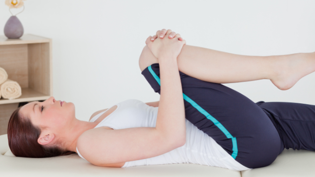
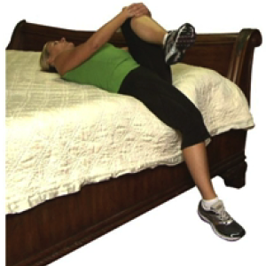
  1. Supine hip flexor stretch
    • Lie flat on your back on a bed with one leg hanging over the side
    • Pull your opposite knee tight toward your chest until a comfortable stretch is felt
    • Hold this stretch for 30 seconds
    • Complete three repetitions on each leg
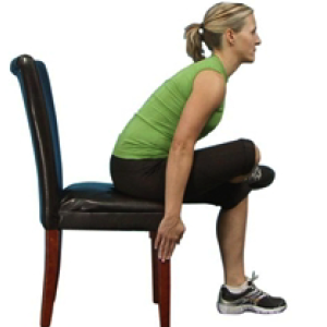
  2. Sitting piriformis stretch
    • While sitting on a chair, cross one leg over your opposite knee
    • Slowly bend your body forward until a comfortable stretch is felt
    • Hold this stretch for 30 seconds
    • Complete three repetitions on each leg
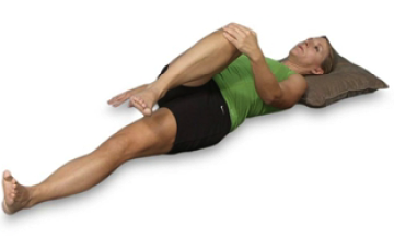
  3. Supine piriformis stretch
    • Lie on your back on the ground
    • Bend one knee up and grab it with your opposite hand
    • Pull your leg across your body, toward your shoulder, until a comfortable stretch is felt
    • Hold this stretch for 30 seconds
    • Complete three repetitions on each leg

Regularly performing these stretches will keep your hips mobile and your risk for hip pain down. But if problems do still manage to arise, it’s best to consult with a physical therapist, who will perform a comprehensive evaluation and design a personalized treatment program that’s suitable for your needs, abilities, and goals.

Hip pain can arise from sudden trauma, overuse, or age-related changes

The hip is a resilient joint that is capable of handling lots of activity and repeated movements, and it can sustain a significant amount of wear and tear. This durability exists because of its complex and multifaceted anatomy that we described in our last newsletter.

But despite these strengths, the hip has a concrete set of limits and is not invulnerable. Repeated activity, not taking enough time to recover, and age can all bring the hip closer to these limits, and when pushed beyond them, the result is injury or pain.

There are a number of conditions that can affect the health of the hip and prevent it from functioning normally, but several problems are seen more frequently due to movement patterns and behaviors that occur in the greater population. Below are 5 of the most common hip-related conditions:

  1. Femoroacetabular impingement
    • Arises when small bony projections (bone spurs) develop along either the femur or acetabulum, causing these bones to rub against each other directly without protection
    • Over time, the resulting friction between these two bones can damage the joint, which will cause symptoms like pain or stiffness in the groin or outside the hip that grows worse with turning, twisting, or squatting motions
  2. Labral tear
    • A rip in the labrum, which covers the acetabulum and secures the femoral head in place
    • The labrum can be torn by sudden trauma or repetitive motion and overuse in sports like hockey, golf, and soccer, age-related changes, structural problems, or some combination of these factors
      • Femoroacetabular impingement can also damage the labrum and lead to a tear over time
    • Symptoms are similar to femoroacetabular impingement and can include pain or stiffness, pain in the buttocks area, a clicking or locking sound in the hip, and feelings of instability
  3. Arthritis
    • Rheumatoid arthritis and osteoarthritis are among the most common causes of hip pain
    • While the course both these conditions take is slightly different, both involve the wearing away of the cartilage that surrounds the ends of bones in the hip joint, resulting in less protection and eventually friction between them as it progresses
    • Symptoms include pain, swelling, tenderness, and stiffness, and a general inability to move the hip and perform routine hip-related activities
  4. Hip bursitis
    • Each hip joint contains two bursae (singular bursa), which are fluid–filled sacs that ease friction between the bones, muscles, and tendons that comprise it
    • Bursitis is the inflammation of one of these bursa, which is typically due to repeated overuse or strain of the hip
    • The primary symptom is a dull, burning pain on the outer hip that’s made worse with excessive walking or climbing stairs
  5. Hip tendinitis
    • Inflammation of any of the tendons that flex the hips
    • Usually caused by repetitive strain and overuse, often from sports or other forms of physical activity
    • Symptoms include pain that develops gradually over time, tenderness, and stiffness in the morning or after long periods of rest

Read our next newsletter for a breakdown of the three most effective exercises that will help you avoid hip pain.

The crucial role the powerful hip joint plays in most body movements

The hip is one of the most versatile and important joints in the body. It’s extremely powerful due to its robust architecture, which also provides it with impressive stability. But the hip joint is also incredibly flexible, allowing for a massive range of motion that is second only to the shoulder in this capacity.

The hip connects the lower extremities to the trunk of the upper body, and its primary function is to bear the body’s weight during both static (eg, standing) and dynamic (eg, walking and running) postures. Bones, cartilage, muscles, ligaments, tendons, and other structures all work together to allow this to occur.

Bones and cartilage The hip is a ball-and-socket joint—the largest in the body—that is comprised of the head of the femur and the acetabulum.

  • Femur: the upper leg bone that connects the knee to the hip; longest and heaviest bone of the body; it has a long shaft and head (femoral head) that serves as the “ball” of the hip joint
  • Acetabulum: a deep, cup-shaped depression of the pelvis bone that serves as the “socket” of the hip joint
  • Cartilage
    • Articular cartilage: the femoral head and acetabulum are covered in articular cartilage at their ends where they meet, which is a strong, slippery, flexible material that allows them to glide smoothly without coming into contact with each other
    • Labrum: a strong piece of cartilage that forms a ring around the outer edge of the acetabulum; it deepens the socket of the pelvis and acts as a gasket to provide the hip with more stability, but it is also elastic to permit greater flexibility

Muscles

Large, thick muscles of the buttocks and thighs surround the hip and are responsible for the significant amount of power that the joint can generate. These muscles are typically classified into four groups—the gluteal group, adductor group, iliopsoas group, and lateral rotator group—based on which directions they permit movement in. The complex anatomy of the hip gives it the ability for three distinct types of movement:

  • Flexion and extension: moves the leg back and forth
  • Abduction and adduction: moves the leg out to the side (abduction) and inward toward the other leg (adduction)
  • Rotation: moves the toes inward (internal rotation) or outward (external rotation)

Ligaments and tendons

The bones and muscles of the hip are all connected by a number of ligaments and tendons that further reinforce the joint’s strength while also preventing each structure from moving too far in any direction. The iliofemoral ligament is the strongest in the body and it prevents excessive adduction and internal rotation, while the pubofemoral ligament restricts abduction and the ischiofemoral ligament prevents internal rotation.

Synovial fluid

Lastly, a synovial membrane encapsulates the entire hip joint, which produces synovial fluid that lubricates and circulates nutrients to the hip. This fluid is stored in the cartilage while at rest but is squeezed out when the hip rotates or bears weight so that it moves more smoothly. As a result, it’s necessary to move the hip frequently to keep it healthy and lubricated.

These structures collectively help to keep the hip strong, stable, and flexible, but as we’ll explore in our next newsletter, problems can occur when any of these components break down or fail to properly perform its function.