Try these 4 exercises for knee mobility and keep your injury risk low

In our last newsletter, we explained why knee pain is so common and explored some of the most common conditions that involve the knee. Knee pain can strike at any age, and while the specific reasons it occurs may vary among different populations, the result is usually the same: an inability to move and function normally in daily life.

We need healthy knees to perform just about any activity that involves the legs, which means the ability to walk, squat, or sit/stand from a chair can be impaired by knee pain. Many of these activities are vital to get through a typical day, meaning that knee pain can prove to be a major hindrance to one’s quality of life. For athletes, knee issues can cause further complications by limiting or preventing play entirely until the pain resolves.

One of the main reasons knee pain occurs so frequently is an overall lack of mobility in the joint, which is often due to inactivity. The good news is that you can improve your knee mobility and reduce your risk for knee pain in the process by performing exercises that target the muscles surrounding the knee. We recommend the following:

Four mobility exercises to reduce your risk for knee pain

Disclaimer – This article and associated images is for educational purposes only. They are not meant to be a substitute for physical therapy or medical care. Please consult with your physical therapist and/or doctor before you start this or any other exercise program.
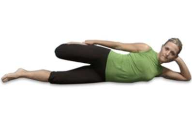
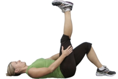
  1. Quadriceps stretch
    • Lie on the floor on one side
    • Grasp your ankle and gently pull your heel up and back until you feel a stretch in the front of your thigh
    • Tighten your stomach muscles to prevent your stomach from sagging outward, and keep your knees close together
    • Hold for about 30 seconds
    • Switch legs and repeat
  2. Hamstring stretch
    • Lie on the floor near the outer corner of a wall or a door frame
    • Raise your left leg and rest your left heel against the wall
    • Keep your left knee slightly bent
    • Gently straighten your left leg until you feel a stretch along the back of your left thigh
    • Hold for about 30 seconds
    • Switch legs and repeat
    • As your flexibility increases, maximize the stretch by gradually scooting yourself closer to the wall or door frame.
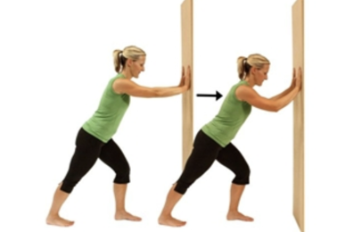
  3. Calf stretch
    • Stand at arm's length from a wall or a piece of sturdy exercise equipment
    • Place your right foot behind your left foot about a foot away
    • Slowly bend your left leg forward, keeping your right knee straight and your right heel on the floor
    • Hold your back straight and your hips forward
    • Don't rotate your feet inward or outward
    • Hold for about 30 seconds
    • Switch legs and repeat
    • To deepen the stretch, slightly bend your right knee as you bend your left leg forward
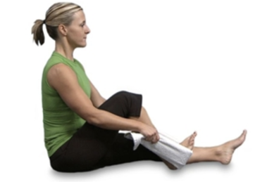
  4. Knee range of motion exercise
    • Sit down with both legs out in front of you
    • Place a towel around your ankle and hold it with both hands
    • Pull the towel and slide your ankle towards your buttocks while keeping your heel on the ground
    • Continue pulling the towel as far as your knee can bend
    • Hold for about 30 seconds
    • Slide your ankle back to the starting position
    • Switch legs and repeat

A physical therapist can help ensure that you’re performing these exercises correctly and provide you with additional exercises to further increase your knee mobility. Contact us today to learn more or schedule an appointment.

In our next newsletter, we’ll discuss the role that strengthening exercises can play in alleviating your knee pain.

Knee pain is common when the joint is pushed beyond its limits

The knee joint is built for durability, which is necessary considering how much action it gets on a daily basis. But it can only withstand so much, and when pushed past its limits, the result is pain and injury. Knee pain is the second most common disorder that affects the body’s movement—behind only back pain—and it’s the leading cause of disability in older adults. But struggles with knee pain are seen across the board, with individuals of all ages and activity levels being affected by it.

The knee is the largest and one of the most complex joints in the body, and its complexity is one of the main reasons it’s so vulnerable to injury. The frequency with which it’s used also plays a significant role. The knee is a hinge joint that’s responsible for bearing weight and allowing the leg to extend and bend back and forth with minimal side-to-side motion. It primarily joins the thighbone (femur) to the shinbone (tibia), but also includes the kneecap (patella) and other lower leg bone (fibula). The patella is a small, triangle-shaped bone that sits in the front of the knee within the quadriceps muscle, and it’s lined with the thickest layer of cartilage in the body because of the massive forces it takes on.

Other important structures of the knee joint include the following:

  • Meniscus: crescent-shaped discs that act as a cushion and shock absorber so that the bones of the knee can move without rubbing against each other; each knee has a medial (inner side, larger) and lateral (outer side, smaller) meniscus
  • Articular cartilage: this thin layer of protective cartilage is found on the femur, top of the tibia, and back of the patella also acts as a shock absorber and helps bones to move smoothly
  • Ligaments: these tough bands of fibrous tissue connect bones and promote their stability by preventing too much motion in any direction
    • Anterior cruciate ligament (ACL): prevents the femur from sliding backward on the tibia and the tibia from sliding forward on the femur
    • Posterior cruciate ligament (PCL): prevents the femur from sliding forward on the tibia and the tibia from sliding backward on the femur
    • Medial collateral ligament (MCL) and lateral collateral ligaments (LCL): these ligaments prevent side-to-side movement of the femur
  • Joint capsule: a membrane bag surrounding the knee that’s filled with a liquid called synovial fluid, which lubricates and nourishes the joint

The likelihood of experiencing different knee conditions changes with age

A variety of issues can affect any of these structures of the knees at any time, but there are certain knee conditions that are seen more frequently in different age groups. For children and adolescents, most cases of knee pain are due to traumatic injuries that typically occur in sports with lots of cutting movements like basketball, football, and soccer. Sprains of the ligaments and strains of the muscles and tendons are most common, but the meniscus, ACL, and other knee ligaments can also be torn when the force exerted on the knee is strong enough.

Many young athletes also have to contend with overuse injuries, which result from performing certain movements over and over, leading to minor damage that accumulates over time and goes on to cause inflammation, irritation, and pain. Osgood-Schlatter disease, in which the area just below the knee becomes inflamed, is a common overuse injury in adolescents involved in sports with lots of running, jumping and/or rapid changes of direction. Other overuse injuries include patellar tendinopathy (jumper’s knee), iliotibial band syndrome, and patellofemoral pain syndrome (runner’s knee).

Later in life, some knee issues are seen less often while other new ones start to emerge. Knee osteoarthritis (OA) accounts for the vast majority of knee pain in older adults, with about 45% individuals expected to deal with it at some point. The condition is so common in this older population because the knee sustains small amounts of damage from everyday use over time and due to the aging process. Knee OA usually leads to pain, stiffness, and swelling that makes it incredibly difficult to walk and move the knees normally.

The risk for traumatic injuries (eg, sprains, strains, and tears) also remains high for adults that stay active in sports and physical activities, and the risk overuse injuries tends to increase with older age because of the gradual breaking down of structures that occurs over time. Common overuse injuries of the knee—some of which were mentioned above—include the following:

  • Patellar tendinopathy (jumper’s knee): results from repeated strain of the patellar tendon that attaches the bottom of the patella to the top of the tibia; symptoms include pain and stiffness at the front or below the patella and/or in the quadriceps, and an ache that typically develops after from exercise
  • Patellofemoral pain syndrome (runner’s knee): involves the patella rubbing against the groove of the femur and accounts for up to 25% of all running injuries; common symptoms are a dull pain behind or around the patella, which may be aggravated by running, squatting, climbing stairs, or sitting
  • Patellar instability: a general term used to describe intermittent pain that comes with the feeling of the patella moving excessively or being unstable; symptoms are pain that’s felt under, around, or most commonly, in front of the patella
  • Iliotibial band syndrome: an injury in which the iliotibial band—which runs from the hip to the top of the tibia—becomes irritated or inflamed from rubbing against the patella; symptoms include pain on the outside of the knee or hip that usually arises after running

In our next newsletter, we look into one of the most effective ways to reduce your risk for experiencing knee pain.

Physical therapists provide a crucial service for preventing falls

Falls are scary, and they can be disabling in both direct and indirect ways. Directly, they often cause injuries that can make it difficult to move and function normally. And then indirectly, they can create a significant fear of falling in many individuals, which in turn leads to less movement and activity which can then further increase the risk for another fall. Any way you slice it, falls can do some serious damage to the lives and independence of older adults.

So if you or someone close to you is in the “at–risk” population for falling, you may very well be interested in taking action to somehow lower the chances. There is a variety of steps one can take to work towards reducing their fall risk, but perhaps the most direct and effective solution is to see a physical therapist for specific guidance.

Physical therapists are experts of human movement that specialize in finding ways to help patients move more effectively and confidently. As such, they are perfectly equipped to identify which older adults are at risk for falls and then guide them through the steps needed to improve their health and modify their lives in ways that will prevent falls from occurring.

From screening, to assessment, to prevention The first step of the fall prevention process is determining whether or not someone is at risk for falling. This is done by an initial screen, which can be given to anyone over the age of 65 and typically consists of three questions:

  1. Have you had 2 or more falls in the last 12 months?
  2. Have you fallen recently?
  3. Do you have any difficulty with walking or balance (the therapist will also examine this briefly to make a determination)?

If the answer to any of these questions is “yes,” then the patient is considered to be in the “high risk for falls” category. From here, a much more thorough assessment is needed, which will include a detailed interview about medications the patient is taking, their fall history, and a physical examination to evaluate balance, strength, mobility, and other factors. This assessment allows the physical therapist to more accurately understand the actual risk for a fall and the impairments present in each patient that need to be targeted.

Based on these findings, the therapist will then create a personalized fall-prevention program to begin right away. Every program is therefore unique according to the patient’s specific impairments and abilities, but research has shown that the best prevention strategies include a variety of different exercises, particularly those that aim to improve balance and strength. As patients repeatedly perform these types of exercises, their reaction times will become more automatic, which will consequently reduce their risk for falls. Part of the program will also involve recommendations for regular physical activity in order to boost fitness levels, which is key for fall prevention.

Lastly, a physical therapist will educate patients and provide specific instructions on how to reduce or eliminate hazards in the home environment and elsewhere. Below are some of the most common tips physical therapists usually provide:

  • Conduct a walkthrough of your home—or have a friend/family member do it—to identify possible hazards that may lead to a fall, then make necessary changes
  • Install handrails on both sides of all stairways, avoid clutter and putting any items on the floor, remove throw rugs and make sure your home is well–lit
  • In bathroom, use nonskid mats, a raised toilet seat and grab bars as needed
  • If you’re supposed to use a walking assistive device, be sure you’re using it properly and at all times, both in and out of the house
  • Get your eyes checked once a year, and get adequate calcium and vitamin D
  • If you’re taking numerous medications, learn the side effects and if there are any interactions that can increase your risk of falling
  • Wear shoes with nonskid soles and consider using Velcro or Spyrolaces
  • Take your time, be patient and ask others for help with difficult, risky tasks

While the power to prevent falls is ultimately in your hands, seeing a physical therapist will be extremely helpful for guiding you and to identify the safest approach to keep you on your feet.

Check out these 4 exercises to help reduce the risk for falls.

If you’re an older adult, a fear of falling may weigh on you every day. This is completely understandable, as falls are the top cause of injury for those over the age of 65. Falls also become more common with each additional year of life that passes due to the increased likelihood of developing other health conditions.

But you should not allow this fear to dominate your life or prevent you from moving. A far more beneficial response is to figure out what you need to do to overcome this fear entirely. And how might this be accomplished? By taking steps to significantly reduce your personal risk for falling.

Falls are usually the results of both environmental hazards–like a loose rug or icy walkway–and personal factors like poor balance or mobility. Reducing or eliminating environmental hazards in your home is one of the most important steps to preventing falls, but it’s only part of the solution, especially when you consider that you can’t control the environment outside of your home. The other major step is to keep yourself mobile and active, which will help you to retain your abilities and address any impairments that might be holding you back.

General physical activity that’s carefully executed is a great start, but to truly lower your fall risk, specific exercises are best. Strength, flexibility, balance, and proprioception (sensing your body’s location relative to other things and controlling its positioning) all tend to decline naturally in older age, so these are the areas that are most crucial to work on. With that in mind, here are the four best exercises designed to strengthen your weaknesses and reduce your risk for falling:

Single–leg stance exercise: improves your balance on each leg, which will in turn help with overall balance

  • Hold on to the back of a chair with both hands
  • Slowly lift one leg off the ground and maintain your balance while standing on one leg for 5 seconds
  • Return to the starting position and repeat 5 times; try to increase the time spent standing on one leg
  • Perform with the opposite leg

Heel–to–toe walk: helps you better maintain your balance while moving and encountering obstacles

  • Position the heel of one foot just in front of the toes of your other foot (your heel and toes should touch or almost (touch)
  • Choose a spot ahead of you and focus on it to keep you steady as you walk
  • Take a step by putting your heel just in front of the toe of your other foot
  • Continue for 20 steps total, then turn around and return
  • Repeat five times

Sit–to–stand exercise (basic):strengthens your leg, core, and back muscles, increases overall mobility, and improves balance

  • Scoot or walk your hips up to the edge of the chair
  • Bring your toes back underneath knees
  • (Optional: use your arms to push off the chair or your knees)
  • Lean forward a little to bring your nose over your toes and push up with your legs to a standing position
  • To sit, bend a little at the knees to push your hips toward the chair and lower your body to a seated position
  • Pause before doing the next repetition
  • Aim for 10 repetitions

Heel raise: strengthens the calf and thigh muscles to improve balance

  • Stand with the back of a chair in front of you
  • Keep your feet 6–8 inches apart, flat on the floor, and parallel to each other
  • Bend your knees slightly so that they are not locked out
  • Elevate your heels to rise on to the balls of your feet; while in motion, use the back of the chair for balance
  • Reverse the motion to the starting position
  • Try to complete at least 2 sets with 10–15 repetitions

Making these exercises a regular part of your routine will build your strength and improve your flexibility, balance, and proprioception. This, in turn, will lead to better overall functioning and will reduce your fall risk. So what are you waiting for?

After a fall, hip fractures pose the biggest threat for seniors

Older adults face a slew of potential health issues as they age, but few are more common or dangerous as falling. One of three adults over the age of 65 will experience a fall every year, making them the leading cause of both fatal and non-fatal injuries in this population.

But what usually happens after a fall, and how does it tend to affect these adults’ lives?

According to Medscape, about 30-50% of falls only result in minor injuries like cuts, scrapes, and bruises that may or may not require a trip to the hospital. But the CDC states that approximately 20% of falls lead to more serious injuries like fractures or trauma to the head that typically send patients straight to the ER.

Aside from death, fractures are by far the most serious consequence of falls. Many bones can be injured from the impact of a fall, but hip fractures occur most frequently and pose the biggest threat to older adults. In the elderly community, an astonishing 95% of hip fractures are caused by falls, and more than 300,000 adults over the age of 65 are hospitalized for one of these injuries every year.

Hip fractures are particularly devastating because of their impact on mobility. After these injuries, many older adults struggle to recover and regain their prior level of function, making them unable to live without the assistance of a caretaker. Surgery is also needed for many patients, which comes with its own set of additional risks. Sadly, as a result, a senior has a 27% chance of dying within one year after a hip fracture.

Other bones that are frequently fractured after falls include:

  • Femur (upper leg bone); these fractures lead to a functional decline in about half of patients
  • Pelvis (hip)
  • Humerus (upper arm bone)
  • Radius or ulna (forearm bones)
  • Spine
  • Foot or ankle bones

The impact of falls on the brain and mind must also be considered

In addition to fractures, falls can also cause head trauma and result in a brain injury, which can be extremely serious. The fall does not need to be severe in order to do damage, and injuries to the head may not be as easy to identify as fractures and other complications. This is why all older adults should visit their doctor right away if they’ve hit their head during a fall to ensure they haven’t suffered from a brain injury.

A final consequence of falling that needs to be understood is the mental toll it can take on each patient. If a fall does occur, many individuals go on to develop an even greater fear of falling, even if they’re not injured. This can cause them to limit their activities, which leads to reduced mobility and loss of physical fitness. Worst of all, this process can turn into a vicious cycle that actually increases the risk for falling because of these changes. The only way to break out of this cycle is to overcome the fear by moving more, building back strength and mobility, and eventually regaining the confidence to continue remaining active.

The facts about falls in the elderly community can be startling

If you or a loved one is over the age of 65, you’re probably aware that there are some dangers associated with falling. There’s no shortage of attention on the topic, with new outlets and experts often discussing the risks involved and offering suggestions on how to prevent falls from occurring. But just how big of a problem are falls in the elderly community, and why do they occur?

Below are a few statistics from the CDC that should help put the matter in perspective:

  • Falls are the leading cause of non—fatal injuries that lead to hospital admissions and death for older adults
  • One in three Americans over the age of 65 will fall at least once every year
  • Every 11 seconds, an older adult is treated in the ER for a fall, and every 19 minutes, an older adult dies because of a fall
    • This equates to approximately 2.8 million visits to the ER, more than 800,000 hospitalizations, and 27,000 deaths each year
  • The death rate associated with falls for seniors increased by 30% from 2007 to 2016
  • In 2015, the total cost of fall—related injuries was about $50 billion
  • Less than half of those who experience a fall tell their doctor about it

Health affects fall risk more than age

So why are falls so common in seniors? Believe it or not, but age itself is not responsible for the increased risk for falling that is said to begin around 65 and increase steadily each additional year. Instead, it all comes down to health, as the chances of experiencing health complications do rise aggressively with advanced age. So even though individuals over the age of 85 are technically considered “high risk,“ an 85—year—old who is in perfectly good health does not necessarily have any greater risk for falling than those in the 65—84 age group.

Once an individual turns 65, a host of health conditions are more likely to occur that can directly affect their chances of falling. Arthritis, dementia, diabetes, vitamin D deficiency, balance impairments, lower body weakness, and impaired vision or hearing are all problems that tend to increase in frequency in this population. Many older adults also take medications for various issues, some of which can affect balance or have other side effects that make it difficult to stay on one’s feet. These are both major factors that place older adults with numerous health conditions and who take medications at an elevated risk for falls. And the more of these risk factors that are present, the higher that individual’s chances for falling.

Environment also plays a major part in fall risk

The final piece of the equation is environment, which can be another significant risk factor for falls. Not taking the proper precautions can leave a home filled with potential hazards that can either cause a fall or fail to prevent one from occurring. Loose rugs, clutter, slippery surfaces, poor lighting, steep or uneven stairs, and a lack of handrails or grab bars are some of the main culprits, but anything that interferes with a person’s ability to navigate their surroundings can be responsible for a fall. These hazards can be commonplace in many homes if no one provides instruction on how to avoid them, which is one reason why up to 50% of falls are due to environmental causes, and about 80% of falls occur in the bathroom.

Collectively, this all shows why education on fall occurrence and guidance on how to eliminate potential hazards in the home are both key to mitigating the risk for falls in older adults.

No matter what is causing your shoulder pain, PT is the best solution

Most people just aren’t themselves when they can’t move properly. This rings especially true for shoulder pain, which is one of the most widespread complaints of the musculoskeletal system that people have.

Some studies estimate that up to 67% of the population will experience shoulder pain at some point in their lives, which makes sense when you think about how frequently we use our shoulders. Any time you reach, lift, push, or pull an object, you’re engaging your shoulder, meaning that they both get used quite frequently on a daily basis.

Shoulder pain can come about from a variety of reasons, but most cases are related to this repeated use that shoulders are put through every day. The shoulders are worked even more aggressively in individuals that do lots of overhead activities as part of their job or sport (like painters, carpenters, swimmers, and baseball and tennis players). Older adults deal with their own set of problems as well, as gradual damage to the structures of the shoulder from repeated use accumulates over time and eventually causes symptoms.

The result of this sustained damage is that a range of shoulder conditions can come about, with the majority involving the rotator cuff, a group of four muscles that surround and stabilize the shoulder joint. Issues like rotator cuff tendinitis—the most common shoulder injury—shoulder impingement, shoulder bursitis, and frozen shoulder each produce a unique set of symptoms, but they all interfere with one’s ability to perform many movements that involve the shoulder normally.

All painful conditions that affect the shoulder share something else in common: the best way to address them is through a course of physical therapy. Physical therapists are movement experts whose goal is to guide patients back to full strength and function with an exercise-based approach. For patients with any type of shoulder condition, a physical therapist will first focus on identifying the source of the pain, and then design a personalized treatment program that targets any areas of weakness or impairment and teaches patients how to regain their abilities through movement.

Typical physical therapy treatment programs for common shoulder conditions

Most treatment programs will involve some combination of pain—relieving interventions, flexibility and strengthening exercises, manual (hands-on) techniques administered by the physical therapist, and education on how to avoid future shoulder issues. But the specific approach used will vary depending on the condition present, its severity, and the patient’s abilities and goals. Below are a some of the interventions typically used for some of the most common shoulder conditions:

  • Shoulder bursitis
    • Stretching exercises like Codman’s pendulum swings and active range of motion exercises
    • Strengthening exercises that target the scapular and core muscles
    • Ultrasound and other pain—relieving modalities
    • Posture education
  • Rotator cuff tendinitis (shoulder tendinitis)
    • Stretching and strengthening exercises, including external and internal rotation, forward flexion shoulder raises, pendulum exercises, and scapular squeezes
    • Education on how to improve posture and avoid habits that will further aggravate the shoulder
  • Shoulder impingement syndrome
    • Stretching exercises
    • Strengthening exercises that target the rotator cuff and scapular muscles
    • Manual (hands-on) therapy, which typically includes soft—tissue massage
  • Rotator cuff tear
    • Passive treatment like ice, heat, and ultrasound to alleviate pain
    • Strengthening exercises that target the pectoral and upper back muscles
    • Education on how to avoid positions and movements that can further aggravate the shoulder, like sleeping on the side and carrying heavy loads
  • Frozen shoulder
    • Treatment for frozen shoulder depends on the current stage of the condition, from stage 1 (pre-freezing) to stage 2 (freezing), stage 3 (frozen), and stage 4 (thawing)
    • The bulk of treatment consists of manual therapy and stretching and strengthening exercises, which increase in intensity with further stages of the condition; activity-specific training is usually added at stage 4
  • Shoulder dislocation
    • Immobilization of the shoulder for a period of time
    • Stretching exercises (both active and passive) at first
    • Strengthening and functional exercises as the shoulder regains mobility
  • Calcific tendinitis
    • Ice and/or heat
    • Ultrasound
    • Stretching and strengthening exercises for the rotator cuff muscles, which can decrease the pressure on the calcium deposits
    • Extracorporeal shock wave therapy, a treatment that administers high frequency sound waves to the shoulder to break up calcium deposits

Many people will shrug off painful shoulder symptoms at first, but if left untreated, it can lead to additional pain, disability, decreased quality of life, time out from work, and frustration. This is why you should see a physical therapist at the first signs of shoulder pain to get on your way to a full recovery.

Keeping your shoulder mobile may reduce your risk for feeling pain

You probably don’t realize how much you rely on your shoulders unless you’ve dealt with an issue that has made it difficult to use it normally. Practically every moment you perform that involves your hands or arms impacts the shoulder or requires it to be completed. The shoulder is essential in most of the activities you do in any given day, and it opens up the possibility of a wide range of functions.

Unfortunately, the shoulder is also one of the most commonly injured joints in the body, which is primarily due to it being so mobile and allowing the arm to move in so many directions. A variety of shoulder conditions can strike at any age, with some problems being more likely to occur later in life due to an accumulation of damage and natural bodily changes. Most cases of shoulder pain are related to the rotator cuff, which is a group of four muscles that stabilize the shoulder and protect it from injury. Although the mechanism behind all shoulder injuries is different, the end result is usually the same: a recurring pain that interferes with one’s ability to reach overhead and perform many activities that involve the shoulder.

But even though shoulder pain is so common, this does not mean it’s not a foregone conclusion, even if you participate in lots of overhead activities. Shoulder pain can be prevented, and the most effective way to keep your risk for injury low is by increasing its mobility and stability with a specific set of exercises.

There are many muscles that contribute to the function of the shoulder, including those of the upper and mid-back, chest, and shoulders, and the more toned and balanced these muscles are, the lower the risk for shoulder injury. Maintaining the strength of these muscles will also help you achieve symmetry and optimize your functional movements that involve the shoulder with better flexibility and mobility. The end result is a reduced risk for all causes of shoulder pain. Below are four of the best exercises to keep your shoulders mobile and strong and prevent injury:

The 4 best shoulder exercises for injury prevention

  • Scaption: this term is an abbreviation for “scapular plane elevation” because it involves the scapula (shoulder blade); scaption exercises employ the scapula and rhomboid muscles of the upper back to strengthen the muscles of the rotator cuff, particularly the supraspinatus, and will also help to keep the shoulder more mobile and flexible
    • How to perform: stand with a weight in one hand and your thumb pointed toward the ceiling; raise your arm at a 45-degree angle from the body toward ceiling to shoulder height for five seconds
    • Repeat for two sets of 10 repetitions, twice per day
  • Bent over row: this exercise strengthens several muscles of the upper back, including the rhomboids, latissimus dorsi, erector spinae, and trapezius; building up your back muscles will prevent your shoulders from rolling forward and will also help to reinforce good principles of shoulder retraction, which is important for healthy shoulders
    • How to perform: bend forward with one arm hanging down while keeping your spine straight; pull your elbow toward the ceiling and squeeze your shoulder blades inward, holding for five seconds
    • Repeat for two sets of 10 repetitions, twice per day
  • Resisted external rotation: this type of exercise primarily targets the infraspinatus, which is a very important rotator cuff muscle; its main function is to rotate the upper arm bone (humerus) away from the body, and keeping this muscle strong and mobile will therefore improve one’s ability to perform this movement and reduce the risk for injury in the process; it also strengthens another rotator cuff muscle called the teres minor and the deltoids of the shoulder
    • How to perform: lie on your side with your head supported by a pillow or hand, and place a towel roll between your elbow and torso; while holding a weight and keeping your arm bent at 90 degrees, move your hand towards the ceiling and hold for five seconds
    • Repeat for two sets of 10 repetitions, twice per day
  • Chest press: the chest press is one of the most effective muscles for the upper body, as it works the pectoral muscles in the chest, the deltoids in the shoulders, and triceps in the arms; maintaining the strength of these muscles will add to the stability of the shoulder to further increase injury protection
    • How to perform: stand facing the wall with your feet shoulder-width apart, about 2-3 feet away from wall; place your hands on the wall at shoulder height, and then slowly bend your elbows and lean forward, then extend your elbows, holding for five seconds
    • Repeat for two sets of 10 repetitions, twice per day

Most cases of shoulder pain are related to the rotator cuff

Shoulder pain is an extremely common complaint. Up to 26% of the population has it to some degree, and it ranks third—behind back pain and knee pain—in musculoskeletal conditions (those involving the bones, muscles, and related structures) that lead people to consult their doctor. About 1% of the population visits a doctor for shoulder pain each year, and while there are a number of possible issues that may be responsible, several similarities tend to be consistent throughout.

The causes of shoulder pain can generally be categorized into two groups: 1) traumatic (acute) injuries that immediately damage certain structures of the shoulder, and 2) overuse injuries, which occur gradually over time due to continuous strain on the shoulder. In both cases, those who are most likely to experience shoulder pain are individuals who regularly perform overhead movements. These types of movements are necessary in professions like painting and construction, and in sports like baseball, swimming, and tennis, making those who are involved in these activities vulnerable to all types of shoulder issues.

Any component of the shoulder can be damaged in an acute or overuse injury, but the majority of shoulder conditions—about 85%—involve the rotator cuff. The rotator cuff is an important group of four muscles that surround the bones of the shoulder. Each of these muscles spans from a different part of the shoulder blade (scapula) to the head of the upper arm bone (humerus) to form a “cuff” that controls and stabilizes the shoulder. A number of issues can affect the rotator cuff and other structures of the shoulder to cause pain;, and a list of the seven most common conditions follows:

7 most common shoulder conditions

  • Shoulder bursitis: a bursa is a fluid-filled sac that acts as a cushion to prevent structures from rubbing against each other; the subacromial bursa in the shoulder is the largest bursa in the body, and when it becomes inflamed—often from regularly performing too many overhead activities—the result is shoulder bursitis; the most common symptom is pain at the top, front, and outside of the shoulder that gets worse with sleeping and overhead activity
  • Rotator cuff tendinitis (shoulder tendinitis): the most common cause of shoulder pain, this condition results from irritation or inflammation of any of the rotator cuff tendons, which occurs gradually over time; the main symptoms are pain and swelling in the front of the shoulder and side of the arm, usually while raising or lowering the arm
  • Shoulder impingement syndrome: a condition in which the bursa or any rotator tendons are trapped (or impinged) by the two main bones of the shoulder—the humerus and a piece of the scapula called the acromion—which is usually due to an outgrowth of bone (bone spur); symptoms include shoulder pain and weakness, and difficulty reaching up behind the back
    • Note: Over time, shoulder impingement can lead to shoulder tendinitis and/or bursitis, and in some cases the names of these conditions are used interchangeably
  • Rotator cuff tear: the result of one of the rotator cuff tendons detaching from the bone, either partially or completely; these injuries can occur either traumatically due to a single incident, or gradually over time, which is usually the case in older patients; the most common symptom is pain during the day and at night, and when lying on the shoulder or lifting or lowering the arm
  • Frozen shoulder: a condition that occurs when scar tissue forms within the shoulder capsule, another structure that helps to keep the shoulder stable; this causes the shoulder capsule to thicken and tighten around the shoulder joint, which means there is less room for the shoulder to move normally, eventually causing it to “freeze;” symptoms include pain and stiffness that makes it difficult or impossible to move the shoulder
  • Shoulder dislocation: an injury in which the ball of the shoulder (humerus) pops out of the socket (glenoid); this is typically due to a forceful motion, and the dislocation can be either partial or complete; symptoms include pain, swelling, and difficulty moving the shoulder
  • Calcific tendinitis: a condition in which small deposits of calcium form within the tendons of the rotator cuff; calcific tendinitis is most commonly seen in individuals between the ages of 30-60 years, and the reasons it occurs are not entirely understood; in most cases it does not cause symptoms, but can lead to severe pain if the calcium deposits get bigger or become inflamed

Shoulder problems become more common as the body changes over time

You have your shoulders to thank for many of the movements required to get through the day. Driving your car, turning doorknobs, shaking hands, and reaching up or across to grab an item are just a few examples of activities that would not be possible without the extreme mobility that the shoulder provides. The shoulder is the only joint that can rotate a full 360°, making it the most mobile and flexible joint in the body, and it’s this significant range of motion that allows you to perform most of the tasks that involve the arms. There is, however, one downside of all this mobility: it makes the shoulder extremely vulnerable to injury.

Often thought of as a single joint, the shoulder is actually an intricate system that consists of four joints, with the acromioclavicular and glenohumeral joints being most important for movement. The acromioclavicular joint is a gliding joint where a part of the shoulder blade (scapula) called the acromion and the collarbone (clavicle) meet, and it allows the transmission of forces from the upper extremity to the clavicle. The glenohumeral joint is what most people think of when discussing the shoulder, and it’s a ball-and-socket in which the ball is the head of the upper arm bone (humerus) and the socket is the glenoid, a shallow cuplike part of the scapula. It’s this joint that allows for the shoulder’s extremely wide range of motion.

Connecting the bones and muscles of the shoulder are a number of ligaments and tendons, plus several other important structures, including the following:

  • Rotator cuff: a group of four muscles that run from the humerus to the scapula; the tendons of these muscles form a “cuff” around the head of the humerus, and all the muscles work together to allow movement and stabilize the shoulder
  • Labrum: a ring of cartilage surrounding the glenoid that creates a deeper socket for the ball to stabilize the joint
  • Deltoid: the largest and strongest muscle of the shoulder, which provides the strength to lift the arm
  • Joint capsule: a fibrous sheath that encloses the structures of the shoulder joint
  • Bursa: a fluid-filled sac that acts as a cushion between tendons and other structures of the shoulder

Why shoulder issues become more common later in life

As mentioned earlier, the cost of the shoulder’s wide range of motion is that it also means a higher risk for injury. Some of this risk is minimized by the many structures—like the rotator cuff—that provide it with protection and stability, but the shoulder can still be easily injured from a variety of causes.

Shoulder pain is essentially any pain that arises in or around the shoulder. It may originate in the joints themselves or from any of the surrounding muscles, ligaments or tendons that make up the shoulder. Shoulder pain can occur at any age from a variety of causes, with younger individuals usually experiencing issues due to sports participation. But the peak incidence of shoulder pain actually occurs between ages 30-50, at which point issues develop because of gradual age-related changes.

With age, the body undergoes a number of significant changes, many of which increase the chances for pain and injury. In the shoulder, the effects of aging can result from the accumulation of many earlier habits, such as previous injury, overuse or underuse or the shoulder, disorders of certain shoulder structures, or any combination of these factors. Changes in circulation, metabolism, nutrition, and physical fitness can also contribute to the development of shoulder pain and dysfunction.

As a result, everyone is at an increased risk for shoulder pain as they get older, whether they were active individuals or not. But this risk is even higher in those who regularly performed lots of overhead activities—like painters, carpenters, swimmers, and baseball players—at any point in their life. Understanding this risk and what shoulder conditions are most likely to occur in older age can help prepare you for what to expect and how to respond if you do notice any shoulder pain.