
Tennis is a great form of physical activity that works out many parts of the body due to its demanding dynamics, but just like every other sport, it also comes with a risk for injury. The most common injury in the sport is called lateral epicondylitis, which is often referred to as tennis elbow. Tennis elbow is a bothersome injury that can significantly interfere with gameplay, but there are several steps you can take to reduce your risk. And if it does occur, physical therapists have you covered.
The lateral epicondyle is a bony bump on the outside of the elbow that serves as an attachment point for several muscles, tendons, and ligaments of the elbow and forearm. When the arm is overworked, a muscle in this region called the extensor carpi radialis brevis (ECRB) gets weakened, which eventually leads to microscopic tears in its tendon, which attaches to the lateral epicondyle. This results in inflammation of the ECRB tendon, which is called lateral epicondylitis, or tennis elbow.
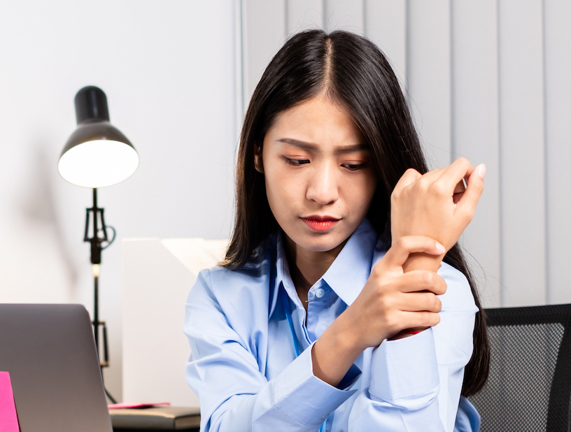
Tennis elbow is yet another example of a repetitive strain injury (RSI) that’s caused by repeatedly performing the same movements in tennis over a long period. Thus, athletes who play tennis and other racquet sports therefore have a particularly high risk for developing tennis elbow, particularly due to the groundstroke in these sports, which directly puts a strain on the ECRB. But tennis elbow can occur in anyone who performs repeated movements that involve the elbow, such as painters, plumbers, and carpenters, who are especially prone to getting tennis elbow. When tennis elbow occurs, the most common symptoms are pain and a burning sensation in the outer part of the forearm and elbow that gets worse with activity, as well as weakened grip strength.
If you play tennis regularly, following these tips can reduce your risk for tennis elbow:
- Learn to use your shoulder and upper arm muscles to take the strain off your elbow
- Stick to the middle of your range of motion during strokes, and avoid bending or straightening your arm all the way
- Make sure your racquet is the right size for you; lighter weight, larger grips, and softer strings may reduce the strain on your tendons
- Take intermittent breaks from tennis throughout the year to avoid overuse
- Try to maintain adequate fitness and flexibility levels with conditioning exercises
- Avoid repeating any one type of stroke, and practice a range of strokes instead
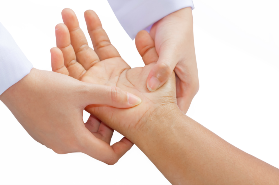
Physical therapy may be needed for patients with bothersome symptoms
But for those of you who are already dealing tennis elbow symptoms, or if symptoms develop in the future, the good news is that 90% of cases will significantly resolve with nonsurgical treatment alone, such as physical therapy. Physical therapists are movement experts who will first perform a thorough evaluation to identify the source of your pain and determine if any of your movements or activities may be contributing factors. From there, the therapist will design a personalized, evidence–based treatment program designed to alleviate your symptoms and restore your physical function with a variety of interventions. A typical physical therapy program for tennis elbow will consist of the following components:
- Elbow bracing, which reduces stress on the ECRB and allows it to self–repair; research has shown that using an elbow brace can significantly reduce the frequency and severity of pain in tennis elbow
- Strengthening exercises that target weakness in the wrist, forearm, and core muscles; eccentric exercises, or negative strengthening exercises, are particularly effective for tennis elbow and are likely to be included
- Manual therapy to increase the flexibility of joints and muscles of the lower arm and alleviate painful symptoms
- Activity modification training, in which the therapist will teach you how to modify any movements you perform regularly that could be contributing to your symptoms
- For tennis players, your therapist may guide you on how to select the right type of racquet, how to modify your stroke to avoid repetitive strain of your, and how frequently you should be taking breaks to avoid overuse
As we’ve shown you over these last four posts, RSIs and occupational overuse syndrome can be the product of a wide range of movements involved in work, sports, or just about anywhere else, and the only real criterion is that the movement is performed repeatedly over a long period. The resulting pain and functional impairments often hold patients back from certain activities and can degrade their quality of life. But on the bright side, physical therapists are ideally suited to identify and treat these injuries with various interventions that both address patients’ symptoms and teach them how to modify their behaviors and prevent future injuries.